Instrumental assessments of swallowing differ from bedside clinical examinations in that they provide objective measures of swallowing function. The gold standard for the instrumental assessment of dysphagia has long been considered to be the videofluoroscopic swallow study (VFSS), also commonly known as a "modified barium swallow." However, there are several other alternative assessment methods that can be considered, especially when the etiology of dysphagia is likely to be esophageal in nature. In addition, many of these assessments are complementary to the VFSS, providing further insight into dysfunction and thus treatment options and prognosis. This article will focus on some practical instrumental swallowing assessment tools, especially those that can be performed in the otolaryngologist's office.
Fiberoptic Endoscopic Evaluation of Swallowing
The Fiberoptic (Flexible) Endoscopic Evaluation of Swallowing (FEES) examination was first described in 1988 as a collaborative effort between otolaryngologists and speech-language pathologists.1 Since that time, it has proven to be a valuable technique in the evaluation of the dysphagic patient. The benefits of FEES include portability, cost effectiveness, and the lack of radiation exposure.
When compared to VFSS, FEES has been found to be more sensitive in the detection of aspiration and penetration.2 In addition, FEES allows detailed evaluation of the mucosal surfaces and the patient's ability to manage their secretions. Drawbacks include the inability to fully evaluate the oral and pharyngoesophageal stages of swallowing. In addition, laryngeal elevation, pharyngeal peristalsis/contraction, and tongue base retraction are better assessed with a VFSS.
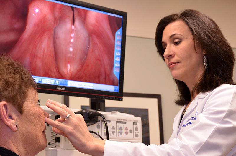
Otolaryngologists are very familiar with endoscopic laryngeal anatomy and often perform flexible laryngoscopy in the initial evaluation of swallowing dysfunction. Therefore, incorporation of a screening FEES examination is not a difficult addition to the otolaryngologist's practice. Performance of the FEES with a speech-language pathologist is preferable, if possible, since a collaborative relationship will lead to enhanced patient outcome and satisfaction. However, swallowing evaluations are within the otolaryngologist's scope of practice and post-graduation training is readily available to give the otolaryngologist the tools needed to perform FEES independently if necessary or desired. The reader is encouraged to see Hiss and Postma (2003), which provides an excellent overview of the FEES procedure.3
FEES is accomplished by passing a flexible laryngoscope trans-nasally. The upper aerodigestive tract is then evaluated while the patient swallows thin liquids (e.g. apple juice, water, or milk), puree (e.g. pudding or applesauce), and a solid (e.g. cookie or graham cracker). Adding a few drops of food coloring can assist in viewing subtle aspiration or penetration into the laryngeal vestibule or more distally into the airway. Any oropharyngeal dysfunction is noted, such as weakness, incoordination, premature bolus spillage, pharyngeal residue, penetration, or aspiration. If any exists, the patient is referred to the speech language pathologist for further evaluation and rehabilitation. If the oropharyngeal examination is normal, then the practitioner can focus efforts on a more thorough esophageal evaluation. For example, pooling of secretions or regurgitation of contents from the post-cricoid region can be indicative of cricopharyngeal dysfunction or high esophageal stricture and warrant further esophageal investigation (Figure 1).
 |
|
Figure 1. FEES examination in a patient with the complaint of "food sticking" in the mid-pharyngeal region. Exam revealed a coordinated, timely swallow and initially no pharyngeal residue. As the exam progressed, however, she began to spontaneously regurgitate material from the post-cricoid region (A). An esophagram was ordered and revealed an obstructive cricopharyngeal bar (B). Endoscopic cricopharyngeal myotomy was performed with complete resolution of symptoms.
|
Esophagram (Barium Swallow)
Although most practitioners agree that an esophageal "follow through" should be performed as part of the VFSS protocol, many do not include this vital piece of information. A formal esophagram differs from the VFSS in that a detailed esophageal evaluation is completed in multiple views (AP, lateral, and often oblique). An esophageal screening as part of a VFSS is typically a limited evaluation of esophageal clearance in a single view (either AP or lateral).
An esophagogram can evaluate for cricopharyngeal dysfunction, diverticuli, web, stricture, and motility disorders. Provocative measures are usually completed to evaluate for gastric reflux. This test should be considered when the history is highly suggestive for esophageal dysphagia (e.g. progressive solid food dysphagia, regurgitation, or sensation of food sticking in chest or throat) and if FEES and/or VFSS are unremarkable.
High Resolution Esophageal Manometry
Before the advent of High Resolution Esophageal Manometry (HRM), evaluation of esophageal physiology (peristalsis, motility, contraction pressures, etc.) required highly specialized training and significant experience to interpret complicated wave signals and perform detailed pull through techniques. Moreover, manometric evaluation of the pharynx was very rare. HRM utilizes a solid state (versus the old water perfusion systems) 4.2 mm diameter catheter with 36 pressure sensors spaced1 cm apart. This catheter is coupled to a software program that produces pressure topography plots instead of line tracings (Figure 2). These pressure plots are fairly intuitive and provide a detailed representation of esophageal physiology and function.
HRM is performed by introducing the catheter through the nasal cavity and passing it into the esophagus while the patient performs free water swallows. The catheter is positioned to span both the upper and lower esophageal sphincters. Ten 5 ml liquid bolus swallows are performed and recorded (Figure 2). A combination impedance-manometry catheter is available which gives additional information regarding esophageal clearance or retention of boluses.
 |
|
Figure 2. High Resolution Esophageal Manometry showing normal esophageal motility.
|
HRM can characterize esophageal motility disorders, gastroesophageal junction morphology (and therefore dysfunction such as hiatal hernia or outflow obstruction) as well as upper esophageal sphincter resting pressure, relaxation and closure.4 Pharyngeal manometry is still in the experimental stages, but holds promise for providing objective information of pharyngeal peristalsis and contraction. It is important to note that while HRM-impedance can characterize esophageal dysfunction, imaging or endoscopy must also be performed to rule out organic causes of esophageal dysphagia.
Trans-Nasal Esophagoscopy
Transnasal esophagoscopy (TNE) is an office-based unsedated examination highly valuable in evaluating for many causes of esophageal dysphagia. These include diverticula, webs, strictures, rings, masses and esophagitis. Esophagitis can occur from inflammatory conditions (e.g. eosinophilic esophagitis), infectious agents (e.g. candidiasis), or as a consequence of gastroesophageal reflux
(Figure 3).
 |
|
Figure 3. TNE in patient with unexplained weight loss and a history of immunosuppression. Main complaint was regurgitation of food after meals. TNE revealed florid candidiasis (A). Patient was treated with fluconazole for 14 days with significant resolution of dysphagic symptoms and improvement in endoscopy findings.
|
History should guide the practitioner as to the appropriateness of esophagoscopy in the work-up of dysphagia. Unless a definitive oropharyngeal source of dysphagia is identified, however, most patients are appropriate for endoscopic screening. This is especially true in those with unexplained weight loss, regurgitation, or progressive solid food dysphagia.
TNE is safe and well tolerated by most patients. The most common side effects are a small nasal vault precluding passage of the scope, self-limited epistaxis and vasovagal events.5,6 Esophageal perforation is extremely rare as a result of an unsedated TNE and is significantly lower than the perforation rate reported with rigid esophagoscopy.7,8
TNE provides excellent visualization of the esophageal mucosa. A recent randomized crossover study found TNE to be highly sensitive (0.98) and specific (1.00) in comparison to traditional sedated endoscopy.9 One limitation with TNE, however, is the difficulty in fully assessing the cricopharyngeal region, especially in patients with stricture or pharyngoesophageal diverticuli.
Conclusion
Although the presence or absence of aspiration is important in the evaluation of swallowing dysfunction, it is imperative to assess for function and physiology. There are several instrumental examinations of swallowing that can be used in addition or as an alternative to the traditional VFSS, especially when the cause of dysphagia is likely to be esophageal, VFSS results are unremarkable, or findings do not explain the patient's symptoms. Careful history review and physical exam can lead the practitioner to choose the correct assessment tool(s) for their patients.
Assistant Professor
Evelyn Trammell Institute for Voice and Swallowing