|
|
MUSC Otolaryngology - Head & Neck Surgery E-Update October 2016
|
|
Colleagues,
Our ENT E-Updates are designed to provide brief, practical, clinical updates in areas where we all struggle in managing our patients. I hope you are finding these newsletters useful. Your feedback or questions about the E-Update articles, your patients, or any other ENT issue are always welcome. Write to us at [email protected] - And please forward this E-Update to your colleagues who may also benefit from sharing the latest ENT topics. As always, your support is deeply appreciated.
Yours sincerely,
Paul R. Lambert, M.D.
Professor and Department Chair
|
Brief Update on Vascular Anomalies
Krishna G. Patel, M.D., Ph.D.
|
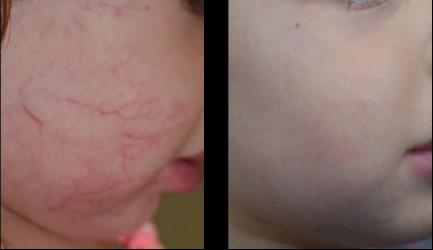
Figure 1.
Multimodal treatment of a hemangioma, initial therapy was propranolol followed by PDL Laser. Left image pre-treatment, Right image post-treatment.
|
The understanding and treatment of vascular anomalies have taken great strides in the past decade. This group of lesions was often confused with each other, and thus, diagnosis and treatments were frequently misdirected. Fortunately, the histopathology of these lesions has become more clearly delineated and better understanding of the cell signaling pathways of these anomalies has led to more directed therapies and improved outcomes.
The organization leading this progress is the International Society for the Study of Vascular Anomalies(ISSVA), founded in 1992 in Amsterdam. The Society promotes both the clinical and research efforts towards better understanding of vascular anomalies such as hemangiomas, venous malformations and lymphatic malformations. As part of this effort, they have continued to refine the classification system of vascular anomalies. Their early classification helped differentiate the tumors, such as hemangiomas, from the vascular malformations such as venous and lymphatic malformations. These differences mostly were defined based upon the fact that hemangiomas exhibit a period of cellular proliferation, where the malformations exhibit normal cell cycle turn over. The classification further differentiated the malformations into low vascular flow lesions, such as lymphatic and venous malformations and the high vascular flow lesions, such as arteriovenous malformations. In 2014, the ISSVA created a much more comprehensive classification that featured the mixed nature of the malformations (
Table 1). For example, frequently these malformations contain more than one cell type, making them a mixed lesion such as lymphatic-venous malformations. Naturally, the dual cell type makes their response to targeted therapies different than if they were purely one cell type.
|
Table 1. Classification of Vascular Anomalies (based from guidelines of the
International Society for the Study of Vascular Anomalies www.issva.org)
|
|
Vascular Anomalies
|
|
|
Vascular Tumors
|
Vascular Malformations
|
|
|
|
Simple
|
Combined
|
of major named vessels
|
associated with other anomalies
|
|
|
Benign Vascular Tumors:
|
Capillary (C) malformations
|
Defined as 2 or more vascular malformations identified in 1 lesion. Can be composed of any combination of: C, L, V, AV*
|
"channel type" or "truncal" malformations
|
Klippel-Trenaunay synd.
|
|
|
Infantile hemangioma
|
Parkes Weber synd.
|
|
|
Congenital hemangioma:
|
Lymphatic (L) malformations
|
Servelle-Martorell synd.
|
|
|
Rapidly involuting (RICH)*
|
|
Sturge- Weber synd.
|
|
|
Non-involving (NICH)
|
Venous (V) malformations
|
|
Limb CM + limb hypertrophy
|
|
|
Partially involuting (PICH)
|
|
|
Maffucci synd.
|
|
|
Tufted angioma
|
Arteriovenous (AV) malformations*
|
|
|
Macrocephaly - CM
|
|
|
Spindle-cell hemangioma
|
|
|
Microcephaly- CM
|
|
|
Epithelioid hemangioma
|
Arteriovenous fistula*
|
|
|
CLOVES synd.
|
|
|
Pyogenic granuloma (aka lobular capillary hemangioma)
|
|
|
Proteus synd.
|
|
|
Others
|
|
|
|
Bannayan-Riley-Ruvalcaba
|
|
|
Locally aggressive or borderline vascular tumors:
|
|
|
|
|
|
|
Kaposiform hemangioendothelioma
|
|
|
|
|
|
|
Retiform hemangioendothelioma
|
|
|
|
|
|
|
Papillary intralymphatic angioendothelioma (PILA),Dabska tumor
|
|
|
|
|
|
|
Composite hemangioendothelioma
|
|
|
|
|
|
|
Kaposi sarcoma
|
|
|
|
|
|
|
Others
|
|
|
|
|
|
|
Malignant vascular tumors:
|
|
|
|
|
|
|
Angiosarcoma
|
|
|
|
|
|
|
Epithelioid hemangioendothelioma
|
|
|
|
|
|
|
Others
|
|
|
|
|
|
|
|
|
|
|
|
|
*
high flow lesions
|
|
|
|
|
|
|
synd. = syndrome
|
|
|
|
|
|
|
M = malformation
|
|
|
|
|
|
|
|
|
|
|
|
|
As the science of these lesions becomes elucidated, management has shifted toward medical therapies over surgical intervention. With respect to infantile hemangiomas, the non-selective beta-blocker, propranolol hydrochloride has become one of the standard and highly successful means of treatment. The successful use of propranolol for treating infantile hemangiomas was first reported in 2008
1, and in 2014, has gained FDA approval under the brand name, Hemangeol (manufacturer: Pierre Fabre Dermatologie). Several level II evidence publications have reported efficacy as high as 97% with propranolol. Less well understood but now making leeway are the vascular malformations. As stated earlier, targeting these lesions can be more challenging because they can be composed of mixed cell types. Sirolimus, a potent immunosuppressant drug, has been under phase trials and when directed toward lymphatic malformations is showing promising outcomes.
Despite the up and coming medical therapies, the current recommended approach for vascular anomalies continues to be the multimodal approach. Depending on the type and properties of the anomaly the following interventions are often combined to optimize treatment outcomes:
- conservative interventions (compression stockings, massage, elevation)
- medical/pharmaceutical management
- laser treatment
- sclerotherapy
- embolization
- surgical excision (partial or complete)
|
|
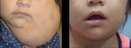
Figure 2. Multimodal treatment of a hemangioma, initial therapy was propranolol followed by surgical debulking. Left image pre-treatment, Right image post-treatment.
|
|
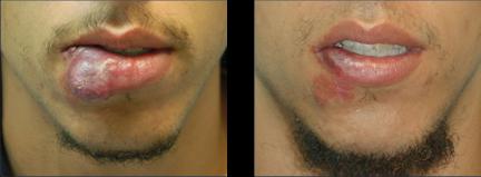
Figure 3. Multimodal treatment of an arteriovenous malformation, initial therapy was embolization followed by partial surgical excision. Left image pre-treatment, Right image post-treatment.
|
|
For example, hemangiomas may be treated initially with propranolol and the residual tumor may be addressed with laser for skin discoloration or surgery for incomplete involution of the tumor (
Figure 1 + 2). Likewise, arteriovenous malformations may undergo embolization for bleeding control followed by surgical excision of the nidus (
Figure 3). No doubt, the advancements in cell signaling will lend to better and more effective medical means of treating these lesions. Until then, the best means of management continues to be a multidisciplinary multimodal approach.
For more information on vascular anomaly classification, go to: www.issva.org
|
References
- Léauté-Labrèze C, Dumas de la Roque E, Hubiche T, et al. Propranolol for severe hemangiomas of infancy. N Engl J Med 2008;358(24):2649-2651.
|
|
Krishna G. Patel, M.D., Ph.D.
Associate Professor and Director,
Facial Plastic & Reconstructive Surgery
M.D. & Ph.D.: Medical College of Georgia
Residency: UNC Chapel Hill
Fellowship: University of California, Davis
Special Interests: Facial plastic and reconstructive surgery, cleft lip and palate repair, facial trauma
Email: [email protected]
|
|
E-Update Articles
Look for these articles in upcoming issues
November:
HPV Associated Oropharyngeal Cancer - 2017 Update
December:
Status of Sustained Release Dexamethasone in Treating Meniere's Disease
January:
Caustic Ingestion in Children
To view any of our past E-Updates visit our
|
|
Continuing Education
November 11, 2016
__________________
2017 Courses:
4th Annual Pediatric ENT Update
February 11, 2017
Temporal Bone Dissection Course
March 24-25, 2017
Southern States Rhinology
March 28-31, 2017
The 17th Annual Charleston Magnolia Conference
June 2-3, 2017
The Charleston Course: 7th Annual Otolaryngology Literature Update
July 14-16, 2017
|
|
|
|
|
|
|