MSA Headquarters Address:
11130 Sunrise Valley Drive, Suite 350
Reston, Virginia 20191
|

M&M 2018
|
|
MEETING UPDATE!
The early registration deadline is fast approaching! Register by June 25 and save $100 on your registration fee!
Don't wait! Book your hotel in Baltimore! Special M&M rates are good through July 1. BOOK HERE.
Want to enter the MICROGRAPH COMPETITION? Click HERE.
Want to share your recent research? Submit your post-deadline paper to present as a poster! Deadline is June 26. Details here.
|
 |
|
Science News
|
|
 The MSA Facebook page
regularly posts science news for you
The MSA Facebook page
regularly posts science news for you
Novel microscopy technique developed to analyze cellular focal adhesion dynamics
Focal adhesions are large specialized proteins that are located in the area where a cell membrane meets the extracellular matrix (ECM), a collection of molecules surrounding the cells that provide support and regulate micromechanical signals to the cells. Examining focal adhesions is one of the key elements to understanding how a cell proliferates, differentiates, and migrates -- which can help in the treatment of diseases like cancer. Read more here.
Team puts the optical microscope under the microscope to achieve atomic accuracy
Over the last two decades, scientists have discovered that the optical microscope can be used to detect, track and image objects much smaller than their traditional limit-about half the wavelength of visible light, or a few hundred nanometers.
Read more
here.
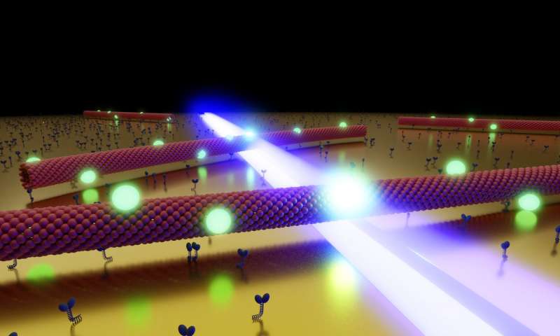
Stage-diving with biomolecules improves optical microscopy
|
|
 |
|
Artistic representation of several microtubules, gliding through the optical near field (blue) of a nanostructured gold surface. The quantum dots (green) attached to the microtubules react to the local field by increasing their fluorescence rate. Credit: Heiko Gross
|
|
Physicists from Dresden and Würzburg have developed a novel method for optical microscopy, obtaining high-resolution images using biological motors and single quantum dots.
The resolution of conventional optical microscopy is limited by the fundamental physical principle of diffraction to about one-half of the wavelength of the light: If the distance between two objects is smaller than this so-called "diffraction limit," they can no longer be visually separated-the image appears "blurred." To acquire optical images at the scale of few nanometers, this is clearly not sufficient. Read more
here.
Don't quite understand how social media works & why it's important? Access this free and interactive
how to guide
.
|
|
|
Association News
|
|

Microscopy Today Digital Editions Available Online
Did you know that you can access past issues of
Microscopy Today
online? You can also view the most cited and most read articles over the past month.
Cryo-EM at 2018 ACA Annual Meeting, Toronto
The cryo-EM community is excited to contribute to the annual meeting of the American Crystallographic Association (ACA) in Toronto, July 20-24, 2018. On Friday July 20, there will be a one-day workshop "Cryo-EM -A Guide to High-Resolution Structure Determination" providing a detailed overview of cryo-EM specimen preparation, image processing and building/refinement of atomic models. Please join us! Registration is now open at
http://www.amercrystalassn.org/2018-meeting-homepage
|
|
In Memoriam
|
|
 It is with deep sadness that we report the passing of Caroline Schooley on April 15, 2018 at the age of 86. Caroline was a graduate of UC Berkeley with a Masters Degree in Microbiology, but began her career in electron microscopy in the early 1950s as an undergraduate, starting as a photography technician in the Electron Microscopy Laboratory and working her way up to becoming director of the EML. In the course of her career she taught countless students the art and science of electron microscopy as both instrumentation and techniques of specimen preparation advanced. She was a strong advocate of collaboration and strongly advised people, especially women, to join the Microscopy Society of America, pointing out it was one of few societies that was degree- and gender-blind.
In addition to being a member of MSA since 1970 and on the Council from 1985-1988, she was an Honorary Fellow of the American Association for the Advancement of Science, a member of the American Society of Cell Biology, the Northern California Society of Electron Microscopy (Secretary 1974-1977), and the Royal Microscopy Society.
Caroline worked on negotiating a microscopy teaching exchange with China for several years. With the help of friends of Mei Lie Wong, it finally happened in May 12 - June 7, 1986. China set up 3 places in various locations in China for 1 week exchanges (Beijing, Xian, and Nanjing). Interested Chinese could sign up, and sign up they did. They came from all over China. Caroline Schooley, Mei Lie Wong and Judy Murphy went on the first exchange. We were thankful to RMC for providing the flights. We were told that the labs were likely not equipped as well as in the US so we should come prepared. We solicited donations from the various microscopy vendors and put together sample prep equipment boxes for each location. We arrived in China with something like 45 boxes, suitcases, etc.
When we arrived in Beijing, there were perhaps 8 people that met us at the airport. After the first location, word spread, and we were met by 20 or 30 people at the following locations to help carry all the materials. After each week's workshop, we raffled all the sample equipment off to the participants. Everyone got something. The Chinese never heard of a raffle before, and they loved it - especially free! It was a unique experience, culturally, and for microscopy. We were all thankful for the warm hospitality and helpfulness of our newly found Chinese friends. We remained in contact with many of those we met, even to this day. All three, Caroline, Judy and Mei Lei said it was absolutely their favorite travel trip. The China exchange continued for many years with Judy Murphy as Director of the China Exchange with two exchanges per year, one biological and one materials oriented. Thank you Caroline for all your efforts in making the exchange happen.
Personal insight
In microscopy, we all know how important networking is and that, it is not just what you know, that is important, BUT who you know that knows what you don't know. Caroline was the queen of networking. If someone needed to know how to do something, or someone to do something, one only needed to lift the phone and call Caroline. She knew EVERYONE, and someone who knew what she didn't know. This went far beyond microscopy, as Caroline had such a creative mind, was an avid reader, and did so many crafty things. She was an artist in her own right. One always saved her Christmas cards, as she personally drew, usually some gorgeous sea creature on the card. She did a lot of tide pooling and had a major exhibit at the light house near Fort Bragg of all the tiny creatures living just under the rocks. She was also a docent for the light house and loved telling folks about its history and of course all the little creatures under the rocks. Before digital photography, she always carried her 110 camera with all the accessories and extra lenses. It is a tiny little camera, with a small film format, but she got some great photos.
In her retirement Caroline helped form Project MICRO, an educational outreach program of MSA, and served as coordinator of the program for many years. The focus of this program is developing interest in science and microscopy among pre-college students. Caroline in particular hoped to inspire young girls into the sciences.
Caroline was part of Project MICRO (
Microscopy
In
Curriculum -
Research
Outreach) from its beginning in 1995. Its goal is to support education about microscopy and the microworld in our schools (principally at the middle school level) nationwide. Caroline was rightly very proud of her involvement with the production of
Microscopic Explorations (1998). This was one of over 70 guides in the GEMS (Great Explorations in Math and Science) series from the Lawrence Hall of Science (LHS), University of California, Berkeley, CA. There was a revised fourth printing in 2007. MSA sponsored the publication of this book, and Caroline was actively involved in its writing. The GEMS program is well known by teachers throughout the country, and has published a series of teachers' guides on activities and similar festivals in different aspects of science. A Spanish translation of the copyable student worksheets is available online (four PDFs) at
http://lhsgems.org/GEMmicro.html Grades 4-8.
She was adamant that Project MICRO should target middle schools. As it turns out, that is exactly where students start to lose interest in the sciences. Microscopy is a beautiful way to bring sciences into the classroom within the comfort zone of the art-based middle school teachers.
She was tireless at the MSA Outreach Booth during M&M conferences, talking to delegates with ideas on how to put ProjectMICRO into effect in their areas. She maintained the junior library often being presented with books by the authors. A lot of her Project MICRO time for the rest of the year was answering emails and helping many with their outreach programs. To give a view of how a program might work, a hands-on workshop was included in the M&M Meeting. In 2005, 34 teachers attended a Project MICRO Microscopic Explorations workshop.
When it became difficult to travel, she encouraged the participation as a co-chair of Elaine Humphrey (2010). Elaine will always be grateful to have Caroline as a co-chair, to guide and ensure deadlines were met and that she didn't have to reinvent the wheel but could call on Caroline's experience at any time. This partnership lasted to date. Caroline was very active in the program up until this year. She kept an eye on the website, updated the book list, developed a brochure for ProjectMICRO, and often sent relevant articles to Elaine.
She wrote several articles for Microscopy Today, for example
Microscopy For Children (2007),
What is ProjectMICRO (2010). In September, 2016, there was a sudden increase in parent and teacher Emails to MICRO asking for further information. She was delighted to find MICRO in the coveted #1 spot when someone did a Google search for "school microscopes."
She was awarded the Morton D. Maser Award in 1992 and became a Fellow of the Microscopy Society of America 2009, cited as "an outstanding microscopist and educator who has made exceptional contributions to microscopy education from K-12 through post-graduate levels."
|
| MSA StC Student Spotlight |
|
In the third of our series spotlighting MSA Student Success Stories, we spent some time learning more about MSA Student Member, Zunlong Ke and his area of research.
Academic background:
I recently graduated from Georgia Institute of Technology with a Ph.D. in Structural Biology. My research focused on the structure and assembly of enveloped virus and utilizing cryo-electron tomography, or cryo-ET. While there, I worked under Professors Elizabeth Wright and Stephen Harvey.
Current research focus:
Recently, I have started a Post-doc program in structural biology at Medical Research Council Laboratory of Molecular Biology in Cambridge, England. I am very excited and will cherish this new opportunity at CMB to continue doing research in structural biology!
MSA involvement:
My first Microscopy and Microanalysis meeting was in 2013 and I really got my feet wet during that conference. As a graduate student, I participated in events which allowed me to get to know other students, postdocs, and faculty in the same field. I really enjoyed the small talks with them. In August of 2014 and July of 2016, I volunteered at conferences for MSA and have continued to be an active member. This year I will keep an eye on the meeting program and stay tuned on twitter for the great talks.
Interesting personal note:
I grew up in China and I came to the United States to pursue the doctoral degree. Everything was new, different, and exciting! During lab meetings, my pronunciation improved significantly with the help of my lovely lab mates; during social events, my behavior was a little bit off at the beginning and was corrected by my roommates and friends. As for the new life in Cambridge, I've never been to the Great Britain before and I'm thrilled!
----------------------------------------------------------------------
MSA Student Bursary Program for M&M 2018
This may be your last chance to take advantage of the MSA student bursary program for the Microscopy & Microanalysis 2018 meeting - spots are filling fast! MSA student members have the opportunity to offset some of the meeting costs by helping with various tasks throughout the week. Many of the MSA Student Council leaders have participated in the student bursary program and enjoyed the networking and camaraderie. If you would like to participate in the student bursary program for 2018 to help offset your meeting costs, send an email to Student Council at the link below. Registration will close soon - don't miss your chance to get involved!
Questions about the bursary/volunteer program or would like to participate contact:
Janet Gbur - Student Council President
------------------------------------------------------------------------
Don't Wait - Save Money at M&M 2018 and Enjoy Benefits of MSA Student Membership!
Did you know MSA Student Members...
- Receive discounted registration to M&M 2018?
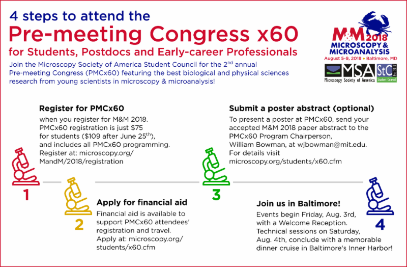
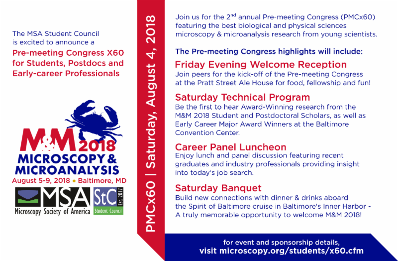
- Receive discounted registration to PMCx60 at M&M 2018?
- Receive Microscopy and Microanalysis and Microscopy Today?
- Can apply for the Student Bursary Program for M&M 2018?
And that's just the start! MSA Student Membership is filled with benefits for undergraduate and graduate students and MSA Student Council programming at M&M 2018 is without a doubt some of the most exciting to date! If you have not done so already, visit "
Join MSA" and renew your MSA Student Membership to take advantage of all the benefits of membership. Let's grow our community!
Remember, M&M Early Bird registration deadline is June 25!
There are multiple ways to connect with the MSA Student Community - Like and follow Student Council on Facebook, Twitter, and Instagram, visit our webpages on
microscopy.org/students or email us at
[email protected].
-------------------------------------------------------------
MSA Student Council - Call for Nominations
MSA Student Council is pleased to announce the Call for Nominations for the executive officer positions. A description of the roles for the three elected positions (President Elect, Treasurer, and Secretary) can be found on the MSA Student Council website,
Responsibilities page. The President-elect serves a three-year term, once each as the President Elect, President, and Past President. The Treasurer and Secretary each serve a one-year term.
Undergraduate and graduate students that are members of MSA are encouraged to run for office and can either self-nominate or nominate other MSA student members. The Call for Nominations letter and Nomination Form can be accessed on the MSA Student Council website,
Responsibilities page and social media platforms. Nominations close on July 29th with elections held at the MSA Student Council Membership meeting during M&M 2018.
-------------------------------------------------------------
|
|
| Local Affiliated Societies |
|
Local Affiliated Societies News
by Patty Jansma, LAS Director
LAS Programs
MSA provides LAS support with Tour Speakers, Grants-in-Aid and Special Meeting grants. Details can be found at
http://www.microscopy.org
/communities/programs.cfm. Funds are still available for 2018. Please contact me as soon as possible if your LAS is interested in applying, and you haven't already received funding this year.
As always, you may contact me at
[email protected] with comments, questions or concerns.
Thanks!
Patty
Microscopy Society of the Ohio River Valley
Ms. Brittany A. Cymes, PhD candidate, Miami University, Oxford, Ohio, received the Student presentation award at the recent Microscopy Society of the Ohio River Valley biannual meeting. Her talk entitled "
Using HR-TEM for Geological Environment Investigation: A Case Study of Silver Nanoparticles Associated with Mine Waste in a World Heritage Site", was presented at the May meeting. The $500 cash award is designed to help defray the cost of attending the 2018 M&M meeting in Baltimore. Congratulations Brittany!
MSORV will hold its next meeting in the fall.
|
|
Focused Interest Group Update
Andrew Vogt, FIG Director
Did you know that there are 11 FIGs that promote a specific discipline to microscopy? Click
here to check them out and contact the leader to get more information. If you are interested in joining one or more of them, please complete the
membership form. You can also join a FIG when renewing your MSA membership; students may join one FIG at no cost.
________________
Focused Interest Group on MicroAnalytical Standards (FIGMAS) Update
 In the summer of 2018, the
Focused Interest Group on MicroAnalytical Standards (FIGMAS) will celebrate its 2-year anniversary. Time for us to look back at the past and onto the future! Our primary goals remain unchanged: building a strong community-based database that would list all available information possible on standards and reference materials used by the microanalytical community (SEM, EPMA, LA-ICP-MS, SIMS, etc.), and preparing the terrane for the reference materials of tomorrow. In the following I would like to update you on news and events around FIGMAS activities.
Web-database
First, we have been working on a web-based interface that would not only list all this information, but also enable users and members of our group to enter new standards information or to suggest an update. This web interface at
http://figmas.org is still in development, and the webmaster (hum... myself) apologizes for taking so long to finish it. I promise to get back to it soon! We also invite you to send your information on standards and reference materials. A forum has also been opened on FIGMAS to allow members to discuss their experience with reference materials, their preparation and maintenance, etc.
Survey and round-robin
Second, we need to evaluate the needs of standards for the next century, and to facilitate the creation of synthetic materials or the (re-)collection of natural ones. A survey has been running over the past month among our members to dress a list of potential candidates for a "new standard", and you will soon have the opportunity to vote for one or more of the suggested materials / minerals. With the help of John Fournelle and Gareth Seward, we are preparing a round robin to permit testing your laboratory performances and evaluating a couple "surprise" minerals... More will be revealed later this year if all goes as planned.
Pre-Meeting Congress at M&M 2018
Let me take here the opportunity to remind you of our
Pre-Meeting Congress X61 at the Microscopy & Microanalysis 2018 meeting in Baltimore (MD) on Saturday August 5th. This PMC, the first of its kind organized by FIGMAS, consists of a one-day meeting combining invited talks, round-table discussions, and poster presentations from contributors. The attendees will learn about and discuss best practices for standard-less and standard-based methods, and for choosing appropriate standards and reference materials for quantitative analysis by EPMA, SEM and other in situ techniques. An overview of speakers and some more information is available at
http://figmas.org. We will also have a
poster session, and everyone is encouraged to show their research on standard compositions, synthesis or sample preparation.
Update on FIGMAS membership
Our list of members has been growing consistently, up to a point where it became the largest Focused Interest Group with 73 paying members from 57 difference academic institution at the end of 2017; 31 members have already renewed their membership in the first couple months of 2018. It confirms the interest (and concerns) of the community about standards and reference materials in terms of quality, availability, material creation, etc. It could also be a side-effect of being the first Focused Interest Group approved and supported by both the Microscopy Society of America and the Microanalysis Society. In any case, this venture would have never been possible without you, and we sincerely thank YOU for your support.
FIGMAS Business Meeting at M&M 2018
We will have again a FIGMAS business meeting at the upcoming M&M meeting. It will take place on Tuesday, June 7th at 12.15 PM. We will inform you about the exact location when we know more.
Upcoming FIGMAS-elections and transition of leadership
At the end of this year, I will leave the leadership of FIGMAS to Anette von der Handt. I am convinced Anette will do an excellent job, and FIGMAS will be in very good hands. I wish her success in this endeavor! On my end, I will remain webmaster of the website and of course in close contact with the community. As a consequence, a ballot will be organized to nominate a new leader-elect and a secretary-treasurer during the FIGMAS business meeting on Tuesday August 8th at noon. Owen Neill is standing for re-election to the secretary-treasurer position, while we are also accepting other nominations. FIGMAS members can nominate a suitable candidate using the form available in the member section of the FIGMAS website (
https://figmas.org/login.php). It has been a great honor and pleasure to meet all of you, and to serve for the community. Long live FIGMAS!
Yours truly,
Julien M. Allaz,
FIGMAS leader 2017-2018
|
|
MSA Job Placement Services
|
|
|
|
|
|
|
|