|
|
Treating Severe Periodontal Disease
And Inflamed Gingival Tissues
|
|
Patient Name: Silver
Age/Breed: 9-year-old FS Mixed Breed
Referred by: Country Side Animal Hospital
|
|
|
History:
Silver is a nine-year-old female, spayed mixed-breed dog referred to The COVE's dentistry department by Country Side Animal Hospital for evaluation and treatment of periodontal disease and inflamed gingival tissues. Silver is also under the care of Virginia Veterinary Centers, for her hypertension, and of Animal Eye Care, for cataracts and glaucoma. Her current oral medications include enalapril and amlodipine, and her ophthalmic medications include atropine, diclofenac and tacrolimus.
Presentation
:
Silver's oral exam revealed generalized severe gingival enlargement. Abrasions were present on the maxillary and mandibular incisors and the gingival enlargement precluded assessment of the maxillary premolars bilaterally on conscious exam. The right enucleation site was well healed. The remainder of her physical examination was unremarkable, including a normal blood pressure of 120mmHg.
Silver's CBC, complete chemistry panel, and UA, completed by the primary care veterinarian, were within normal limits, and Silver was scheduled for anesthesia for a complete oral examination, including intra-oral radiographs, and gingivectomies over the affected areas.
Diagnostics, Diagnosis, and Treatment:
An anesthetized oral exam and full mouth radiographs were performed. This revealed gingival enlargement with corresponding pseudo-pockets affecting the bilateral maxillary incisors; bilateral maxillary canines; bilateral maxillary first, second, third, and fourth premolars; bilateral maxillary first molars; bilateral mandibular; bilateral mandibular canines; and bilateral mandibular first, second, third, and fourth premolars. A complicated crown fracture of the right maxillary second incisor was also found. Nerve blocks were performed at bilateral infraorbital and bilateral inferior alveolar sites, and thorough supra- and sub-gingival scaling and polishing was performed.
Gingivectomies were performed on all the affected teeth, using sharp dissection with a #15 surgical blade, followed by contouring of the gingiva with a #12 fluted bur, taking care to recreate normal beveled gingival margins. While some practitioners use laser or electrocautery to perform the gingivectomy surgery, such techniques risk heat damage to the tooth and thermal necrosis to the underlying bone.
The fractured maxillary incisor was nonsurgically extracted and the gingiva was closed with 4-0 Monocryl, following alveoloplasty.
Recovery was uneventful and Silver was discharged later that day on pain medication.
|
|
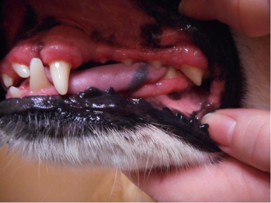
| Pre-operative photo of gingival enlargement |
|
|
|
|
Immediately following surgical gingivectomies
|
|
|
Discussion:
With generalized gingival enlargement, the gingival tissue grows thicker and may cover part of the crown, most commonly the mandibular incisors. This overgrowth creates pseudopockets that result in increased plaque and calculus formation, thereby worsening periodontal disease. Therefore, it is imperative to recreate normal physiologic contours, and two millimeters of attached gingiva, when performing gingivoplasty, and to maintain a diligent home care plan in order to delay recurrence of the overgrowth.
Gingival enlargement is a blanket term that covers both
gingival hyperplasia, the pathological growth of redundant gingival tissue as a result of an increase in the number of cells, and
gingival overgrowth, the increase in gingival volume due to excessive accumulation of extracellular matrix proteins. It is important to remember that neoplasia can have a similar appearance, so histopathology is indicated in these cases.
Certain breeds, including the collie, Great Dane, boxer, and Doberman, are predisposed to develop gingival hyperplasia. In addition, some medications that alter the matrix protein formation have been associated with gingival overgrowth. The drugs associated with gingival overgrowth include immunosuppressants, anticonvulsants, ACE inhibitors, amlodipine, and calcium channel blockers.
In Silver's case, the gingival enlargement is thought to be secondary to medications, especially given the relative lack of periodontal disease on radiographs. Diligent home care may slow recurrence of the disease, but a relapse is expected, unless a medication change can be implemented.
Outcome and Follow-up:
At her two-week recheck appointment, Silver's surgical sites are well healed and her family reports that she is eating well and seems comfortable. They were instructed to initiate daily home care and discuss the possibility of changing from amlodipine and enalapril to alternative medications with their internist. If Silver's blood pressure is unable to be controlled on an alternate medication, frequent recheck examinations and intermittent gingivectomies will be required.
|
|
| Silver at two-week recheck exam |
|
|
|
|
|
TECH TIP: A "How-To" Guide For Periodontal Scoring
-
By Danielle Martin, LVT
|
|
|
|
|
Part of a comprehensive oral health assessment is periodontal scoring of each individual tooth. The periodontal score is the percentage of bone loss that is used to help determine whether extraction is indicated and to keep track of bone loss progressing or improving (with treatment) over time. This should be documented and kept as part of the patient's medical record. Once a technician becomes proficient at scoring, it can be done quickly and will aid the veterinarian in making the most accurate recommendations for treatment and/or extractions. Below are step-by-step instructions for how to perform periodontal scoring. Page references below can be found on the Periodontal Scoring document
here
.
|
- Obtain a diagnostic intraoral radiograph. This must include the entire area from the apex of the tooth to the neck of the crown. Keep in mind that elongating or foreshortening the teeth will not give you accurate measurements, and will therefore cause your periodontal score to be inaccurate.
- Using either your radiograph software system or a ruler to the screen, measure from the neck of the crown (where the bone should be) to the apex of the tooth. This is measurement A. (Page 3)
- Now measure from the shortest level of the bone to the apex of the tooth. This is measurement B. (Page 4)
- Subtract B from A. Example: 7.2 (A) - 4.3 (B) = 2.9 (C)
- C is then divided by A. Example: 2.9 (C) / 7.2 (A) = 0.40 (periodontal score)
- The number you obtained is your periodontal score for that tooth. Example: 0.40 = 40% (Page 5)
Periodontal Disease Classification
- Normal: No gingivitis or attachment loss.
- Stage 1: Gingivitis only with no attachment loss. (Page 6)
- Stage 2: Less than 25 percent bone loss or stage 1 furcation involvement in multi-rooted teeth. (Page 7)
- Stage 3: 25-50 percent bone loss or stage 2 furcation involvement in multi-rooted teeth. (Page 8)
- Stage 4: Greater than 50 percent bone loss or stage 3 furcation involvement in multi-rooted teeth. (Page 9)
Need additional guidance? Consider contacting Danielle Russ at [email protected] or phoning 757.935.9111 to schedule a Lunch and Learn presentation with our dentistry-licensed veterinary technician.
|
|
|
|
COVE News
|
|
COVE News
|
|
|
|
|
As part of our dedication to the veterinarian community, we offer a variety of continuing education opportunities for DVMs, LVTs, and support staff.
Our Lunch and Learns are complimentary educational sessions at your practice during your lunchtime.
Presentations are given by a COVE team member and are approximately 30 minutes to 1 hour in length. You choose the topic and we'll bring the education and food to you!
Some of our most common Lunch and Learn topics are:
- Anesthesia & Analgesia
- Bandaging/Splinting
- CPR
- Dental Emergencies
- ECG Review
- Echocardiography
- ER Triage
- Hypertension
- History and Physical Examination
- Radiographs (dental, thoracic and/or orthopedic)
If you don't see a topic of interest, let us know what you would like to learn about. We'll do our best to accommodate.
To schedule a Lunch and Learn, please contact Danielle Russ, Hospital Manager, with your preferred topic and date
at
[email protected]
or 757.935.9111.
|
The COVE offers Minimally Invasive Surgery (MIS)
In cases where surgery is required, we always look for options that will cause the least amount of pain and provide the quickest recovery option for our patients. In a minimally invasive procedure, small incisions are made and used as passageways for a
laparoscope
or
endoscope
, which are tiny fiber optic video cameras. Working from the images provided from the scope, special instruments are then passed through other openings and operated by remote control to perform the necessary procedure.
Benefits to your patients:
- Smaller incisions
- Quicker recovery time
- Less pain
- Less scarring
- Lower risk of infection
- Reduced blood loss
We offer MIS for:
- Laparoscopy: Abdominal and pelvic surgery
- Thoracoscopy:
Lung/chest surgery
- Arthroscopy: Joint surgery
Would you like to learn more?
Please call us anytime!
|
If you missed hearing our practice manager, Danielle Russ, speak at CVC in December, then you can
check her out at VMX (formerly NAVC) in Orlando in February 2018!
We know you'll enjoy her presentations on:
- Feline Heartworm Disease: the H.A.R.D. part
- Blood Pressure Monitoring in practice
- ECGs: Minding your Ps and Qs
- 7 Deadly Sins of Vet Tech turned Manager
Click
here for registration information.
|
|
|
24/7 Emergency and Critical Care | Surgery | Cardiology | Dentistry
|
6550 Hampton Roads Pkwy, #113 | Suffolk, VA 23435
P: 757.935.9111 | F: 757.935.9110 |
thecovevets.com
|
 |
|
|
|
|
|
|