| |
| |
|
Fall Newsletter
September 2016
|
Wednesday
October 26, 2016
7 - 9 PM
"The Nuts and Bolts of New Horse Ownership"
Presented by
Dr. Joan Ayers
Congratulations on becoming a new horse owner! Come learn about stabling, nutrition, and preventative medicine from a veterinarian's perspective. This course will be most helpful for new horse owners, but those with all levels of experience are welcome to attend. There will be time for a question and answer period at the end along with light refreshments.
_____________ |
|
Don't forget, fall means it's time to do fecal testing for intestinal parasites!

Prepare your sample for your fall vet visit or bring it to the clinic. Collect just a few manure balls, place them in a clean container or zip lock bag, and label with your name and your horse's name. The sample should be no more than 24 hours old and should not have been frozen. Our veterinary technicians Kellie and Emma will be happy to answer your questions about fecals if you call our office at 585-889-1170


_____________ |
|
|
 What's That Bump?
It is not uncommon to find lumps and bumps on a horse's skin. But should you worry? When should you call the veterinarian? A hard-and-fast rule is that the lumps should be monitored carefully and if they grow or change in appearance (lose hair, ulcerate, or drain) a veterinary examination should be scheduled.
Lesions on the eyelids should be examined by a veterinarian sooner rather than later as large masses in this area can lead to loss of the eye. Lumps that are stable in size can be checked by your veterinarian during the annual health assessment exam.
The most common causes of skin masses include collagen granulomas, proud flesh, cysts, and cancer.
|
|
Collagen Granulomas
 |
|
|
|
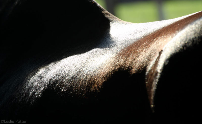
A horse with multiple collagen granulomas on the withers. This is a very typical location for this type of skin bump.
|
Collagen granulomas
are small hard bumps usually found on the horse's back or neck. These bumps consist of scar tissue that forms due to inflammation caused by pressure from the saddle or by insect bites. Diagnosis is most often made by observing the appearance of the lesions and their presence at a typical location on the horse's body. Some horses may be bothered by pressure over the granulomas, which can make them resent being tacked. Discomfort is an indication for treatment, which consists of local injections of a corticosteroid by your veterinarian and by avoiding pressure on the area. Some horses may need injections repeated periodically to keep collagen granulomas under control.
|
|
|
Cysts
Cysts
are usually solitary small nodules covered by healthy skin. Cysts have a hollow center, but can be firm to the touch. They are benign, but they can be difficult to differentiate from other nodules based only on external appearance. Treatment is usually not necessary, and they tend not to recur if surgically removed for histology (examination of cells under a microscope).
|
|
|
Proud Flesh
|
|
|
Proud flesh on the lower hind leg. |
Proud flesh
is excessive granulation tissue that can occur on a healing wound. Horses are known for their propensity to develop proud flesh, especially on wounds on the lower limbs. Diagnosis is made by examining the appearance of the wound. Proud flesh can be treated with topical creams and powders, by using a silicone bandage, or by surgical debridement. Standing wraps or bandages are often needed in order to keep proud flesh under control.
|
|
|
Skin Cancers
|
|
|
The melanoma on the corner of this mare's mouth is currently not causing her any trouble.
|
The most concerning cause of skin bumps is cancer (neoplasia). Melanomas, sarcoids, and squamous cell carcinomas are the most frequently encountered types of skin cancer. Diagnosis can be made by your veterinarian based on the appearance of the lesion and the histopathology results. The histopathology sample can be obtained by either removing the entire lump or by a taking part of the mass. Removing the entire lump is preferable, as it can also serve as treatment, but this is not always possible on large masses. That is another reason to have your veterinarian examine nodules before they get too large. Histopathology not only is helpful to confirm the type of cancer, but also gives information on how aggressive that tumor is.
Melanomas
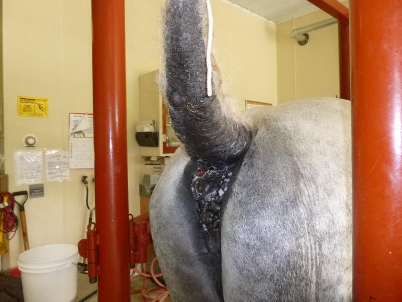
can affect up to 80% of gray horses as they age, but can also occur in horses of other colors. Common locations include the underside of the tail, perineum, around the anus, sheath, throat latch area, and the corners of the mouth.
|
|
|
11 year old gray Percheron mare with large melanomas on the tail, anus, and perineum. These masses are large and extensive, making surgical removal impossible. Her melanomas rupture and drain periodically, attracting flies.
|
These tumors are usually slow growing and may cause no clinical signs in the majority of horses, but they are not benign. They can cause a lot of trouble locally and may metastasize to internal organs. Some complications of local growth include the inability to pass manure due to large masses next to the anus, disruption of function of nearby nerves, and drainage that can become infected and attract flies.
|
|
|
A large single melanoma on the tail of a 17 year old bay pony. Sadly, this mass metastasized to his lung despite tail amputation and chemotherapy.
|
Metastasis
(spreading to another organ) can occur in a small number of horses and does have serious consequences. Treatment options available include surgical removal, chemotherapy, and radiation. A vaccine is also being studied that may help horses with multiple melanomas.
A sarcoid is a type of skin cancer that can cause problems from local growth, but luckily sarcoids do not metastasize to internal organs.
|
|
|
Verrucous sarcoid on a horses's ear.
|
There are multiple factors
involved in the development of sarcoids, including genetic predisposition and the presence of bovine papillomavirus. This kind of tumor occurs more often on the head (including eyelids and ears), neck, inner thigh,
shoulder, legs, and sheath
.
Sarcoids are classified based on their appearance: occult (flat, no hair), verrucous (crusted and wart like), nodular (round masses under haired skin), and fibroblastic (ulcerated
masses).
|
|
|
|
Flat sarcoid on the outside of a hind fetlock that responded well to treatment with imiquimod ointment.
|
|
There are several different treatment options, but no treatment protocol has been universally successful. Options include surgical removal, local chemotherapy, application of topical agents, cryotherapy, and radiotherapy. Recurrence of these tumors after treatment is common. Your veterinarian might recommend benign neglect for smaller sarcoids that are not rapidly changing in appearance. However, there is no guarantee that the lesion will stay harmless.
|
|
|
|
Nodular sarcoids on the inner thigh of a 22 year old mare. These masses had been there for several years and were quiet (pictured on the left), but recently started to grow and ulcerate (pictured on the right).
|
|

Squamous cell carcinoma
is more common in pink skin and it is thought to be associated with sunlight exposure. Sites most commonly affected include eyelids, muzzle, sheath, penis, and vulva. This type of cancer can spread to
internal organs.
|
|
|
|
The muzzle of this 9 year old draft horse has several small lesions that were confirmed by biopsy to be squamous cell carcinoma. His third eyelids were also suspected to be affected.
|
|
Treatment options include surgical removal, local chemotherapy, cryotherapy and radiotherapy. Applying a long fly mask with high UV protection is recommended for horses with white/pink hued eyelids and muzzle in order to prevent this kind of tumor.
|
|
|
|
Minor ulceration on white eyelids like this can be an early sign of squamous cell carcinoma and should be examined by a veterinarian. |
When dealing with skin cancer it is always better to address the lesions at an early stage when they are still small and less likely to cause destruction of nearby tissues or to spread to internal organs. There are many conditions that cause skin bumps, however, and evaluation by your veterinarian is crucial in order to decide if additional tests or treatment is required.
|
|
| |
| |
| |
|