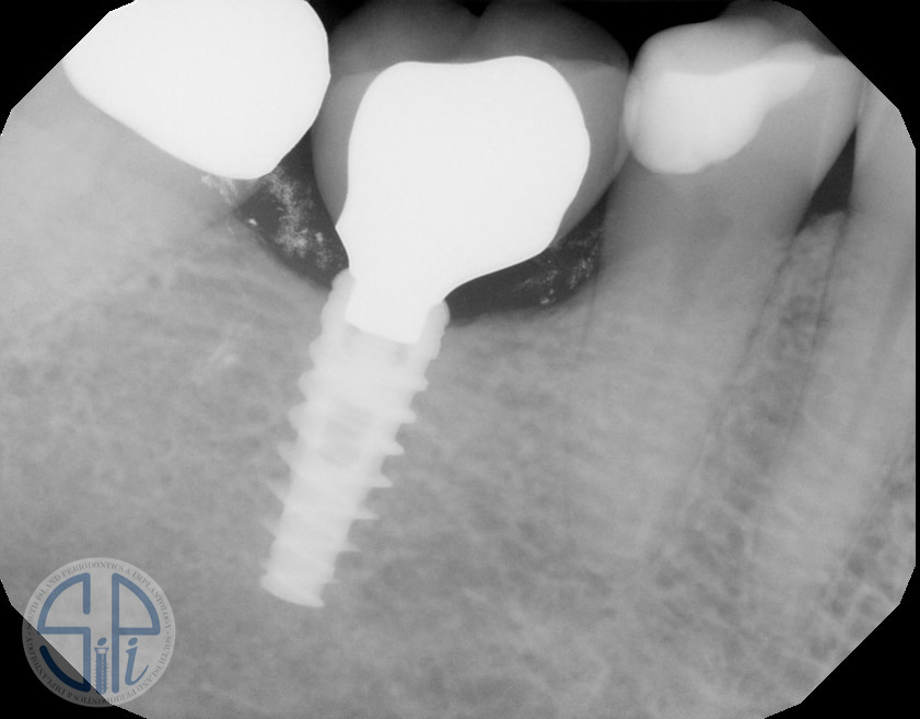
In 2020, seven years after initial restoration, a periapical radiograph revealed stable bone levels around implant #30.

Tooth #29 was restored with a full coverage crown secondary to root canal therapy. An open contact was present between #29 PFM and implant crown #30.
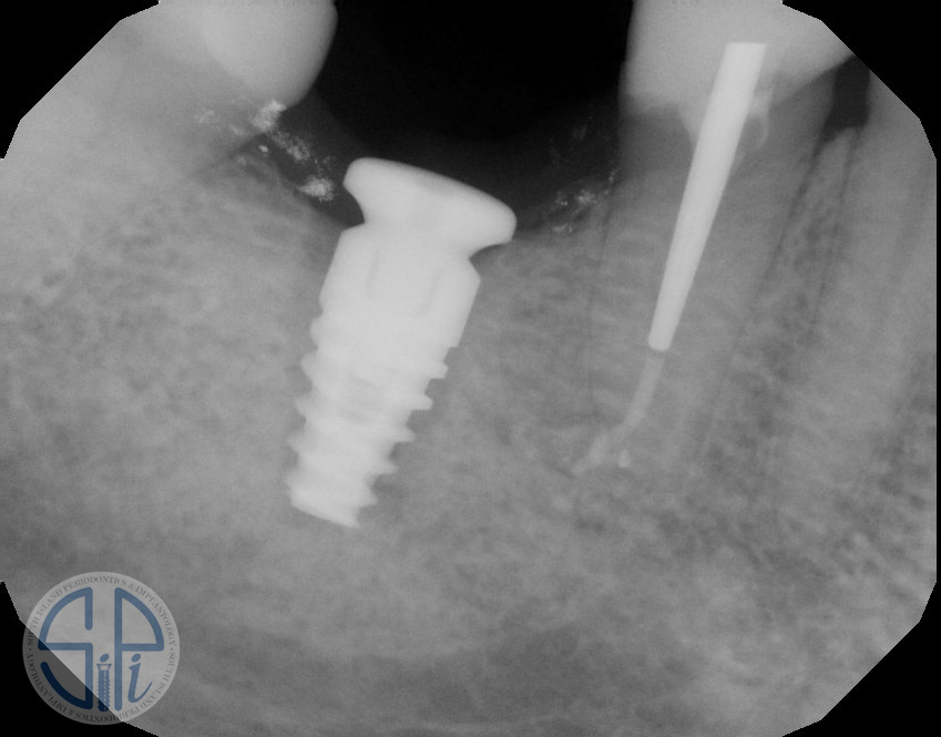
Last month, the patient presented with a clinically mobile implant. The updated periapical radiograph showed a radiolucency along the entire implant. The implant failed and had to be removed.
What could possibly account for this rapid progression of bone loss? If we take a closer look at our radiograph we will notice that there is a new post and crown on adjacent tooth #29 that was replaced within the last year. The contact persistently remains open. Open contacts adjacent to implant restorations are common and cause food traps and subsequent caries on proximal teeth.

The occlusal scheme, or lack of one, of the new restoration appears to have overloaded the implant and caused rapid breakdown of bone and osseointegration of the implant.

At this point, the patient was informed that the implant had a hopeless prognosis, and she consented to implant removal and replacement.

Integrated implant #30 likely failed because the occlusal forces on implant crown #30 were not controlled when tooth #29 received its most recent restoration. The new crown on tooth #29 could have caused posterior balancing side interferences in excursions on implant crown #30 which ultimately caused bone loss and implant failure.
South Island Periodontics and Implantology | SiPi
‌