I-site newsletter is a global educational newsletter FOR HEALTH CARE PROVIDERS launched in 2009, that monthly provides independent updates from the international literature on specialty rigid contact lenses such as corneal, orthokeratology and scleral lenses and related topics. View as Webpage
|
|
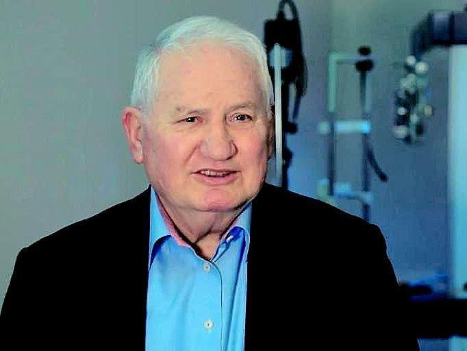
An icon in the scleral lens field sadly passed away, today exactly one month ago. Rients Visser was one of the original pioneers of scleral lenses, and the name Visser is renowned worldwide in the field. He was one of the few in the contact lens community who, about 40 years ago, kept believing in scleral lenses. Despite the criticism from a number of opponents - pointing at the lack of lens movement with sclerals, the theoretical limited oxygen delivery to the cornea, the potential intraocular pressure increase, etc. - Rients simply kept saying: 'but Eef, it works! And I can make patients happy'. He urged us to continue the path of fitting sclerals to patients who could benefit from them, while finding scientific answers to the questions raised. Rients was perhaps one of the first in the world to recognize the benefits related to using toric-back-surface scleral lenses and did this without having access to proper imaging technology, and with that he was ahead of his time in acknowledging the irregular nature of the scleral shape. Toric and other asymmetric scleral lenses have become mainstream now, but Rients Visser for decades started with a toric scleral lens as the first lens option for every patient. If in some rare cases the sclera happened to be spherical, he could always return to a spherical back surface. But in the majority of eyes, that was not the case. Rients received the Dutch Jan-Kok penning in 2015 at the bi-annual optometric conference OVN and received the 'Global Specialty Lens Symposium Award of Excellence' in Las Vegas in the same year. Rients passed away just days after his 80th birthday in his hometown of Nijmegen in the Netherlands and in the presence of his wife, children and grandchildren. Although Rients is not with us anymore, he will always be linked to the development of scleral lenses and remembered as not only a visioneer but also as a very cordial and warm-hearted person.
Eef van der Worp
|
|
Differentiating Between Contact Lens Warpage and Keratoconus Using OCT |
|
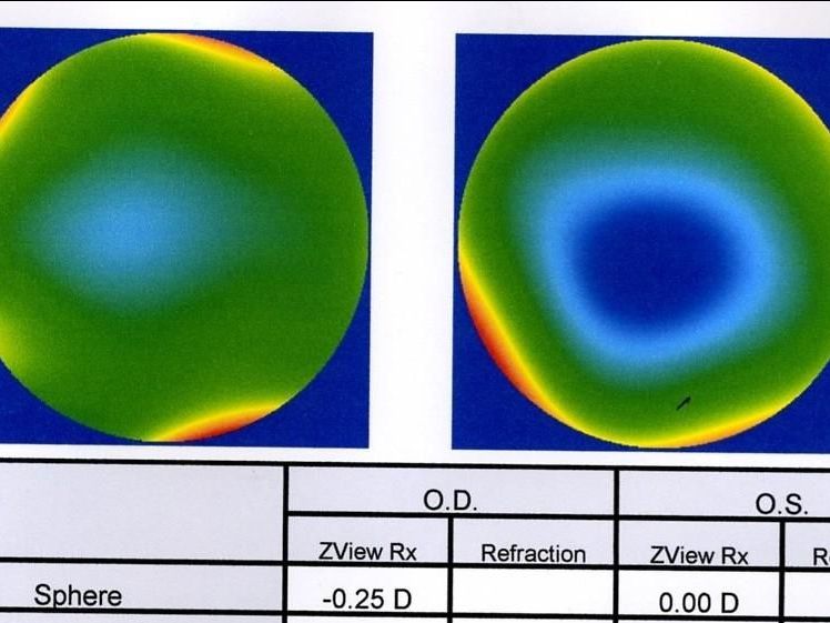
Normal eyes and eyes with either contact lens warpage or keratoconus were scanned by a Fourier-domain optical coherence tomography (OCT) system. Maps of epithelial thickness and anterior surface mean curvature were generated and converted to deviation maps by subtracting the average maps from a healthy population. The average Epithelial Modulation Index (EMI) value for normal eyes was -0.6±1.0 µm/m. Eyes with keratoconus were characterized by coincident high anterior surface mean curvature and low epithelial thickness, resulting in a high EMI index (manifest keratoconus 103.0±82.9µm/m, subclinical keratoconus 37.0±23.0µm/m and forme fruste keratoconus 7.3±13.2µm/m). The EMI index was closer to normal for eyes with warpage (-1.9±4.0µm/m). The classification accuracy of the index was 100±0% for normal eyes and 99.0±2.0% for eyes with warpage, while it was was 100±0%, 100±0% and 53.1±1.5% for the manifest, subclinical, and forme fruste keratoconus groups, respectively. In conclusion, the investigators believe that OCT and this index are useful in distinguishing eyes with keratoconus from those with contact lens-related corneal warpage. Image: Gustav Poltner
|
|
Higher-Order Aberrations in Different Stages of Keratoconus |
|
Researchers from Japan measured higher-order aberrations (HOAs) in clinical and subclinical keratoconus (KC). The study included 33, 36, and 26 patients with clinical, topographic (no clinical signs), and pre-topographic (normal topography and no clinical signs) KC and 30 controls without KC. Control eyes and eyes with pre-topographic KC had significantly lower ocular or corneal total HOAs and Zernike vector terms than those with clinical KC and topographic KC, except for ocular tetrafoil between topographic KC and pre-topographic KC and spherical aberration among all groups. The found difference in corneal total HOAs and corneal coma in this study, could serve as a potential tool to discriminate pre-topographic KC from normal control eyes. |
|
Peripheral Refraction and Higher-Order Aberrations in Ortho-k vs Soft MF Lenses |
|
This study from China compared the peripheral defocus, higher-order aberrations (HOAs) and contrast visual acuity in myopic children wearing ortho-k (OK) lenses and multifocal soft contact lenses (MSCLs) designed with high addition. Myopes between 1.00-5.00D and between 8-13 years of age were included. After wearing the lenses, subjects in the MSCL group had relative peripheral corneal defocus and relative peripheral refraction values similar to those in the OK group at the paracentral retina (within 2 mm of the cornea or 20° of the retina) but larger than those in the OK group at the periphery. All HOAs increased after wearing the lenses except for the trefoil in the MSCL group. HOAs increased more in the OK group. The 100% and 10% contrast acuities were worse in the MSCL group. After one year, axial elongation was 0.37mm (SD=0.16) in the MSCL group and 0.28mm (0.16) in the OK group. In conclusion: MSCLs produced larger myopic defocus at the periphery, increased HOAs less and resulted in worse contrast acuity compared to OK lenses. The high addition of this MSCL did not result in better myopia control efficacy. |
|
Eyelids & Tears in Scleral Lens Wear |
|
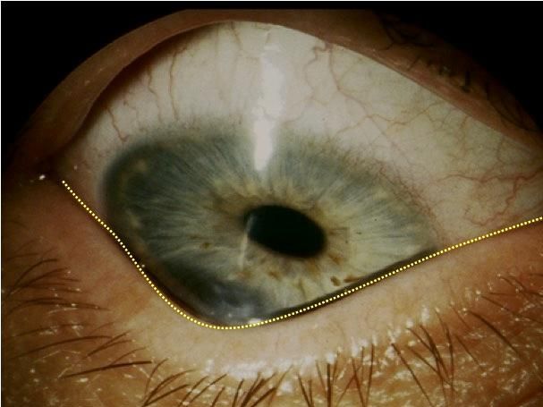
One of those questions that remained unanswered for a long time was: What are the inflammatory consequences of wearing scleral lenses? Maria Walker and co-workers shed some important light on this in this review article. Scleral lenses can affect a range of anterior segment structures including the eyelids and also the tear film. The eyelids, consisting of the outer skin layer, the middle tarsal plate and the posterior palpebral conjunctiva, provide physical protection and house the meibomian glands and cilia, which have important and unique functions. Tears consist of a mix of aqueous, mucus and lipidomic components that serve vital functions of lubricity, protection and nourishment to the ocular surface. Both the eyelids and the tear film interact directly with scleral lenses on the eye and can affect but also be impacted by scleral lens wear. This paper provides a review of the anatomy and physiology of the eyelids and tear film, discusses the effects and impacts of the scleral lenses on these structures and identifies areas that require further research.
|
|
In some earlier work, Walker et al in Contact Lens & Anterior Eye measured inflammatory mediators in the scleral lens fluid reservoir (FR) in healthy eyes and compared that to basal tear samples. Fifteen normal, habitual soft contact lens wearers were fitted with 14.8mm or 15.4mm scleral lenses, and basal ocular surface tears and FR samples were collected. Levels of interleukin (IL) -4 and -8, matrix metalloproteinase (MMP)-7, -9 and -10 and tissue inhibitor of MMPs (TIMPs) 1-4 were collected. The MMP-9 and MMP-10 values were greater in the FR than in the basal ocular surface tears. After 8h of scleral lens (SL) wear, the median concentration of MMP-9 in the FR and basal tears were 62.7ng/ML and 15.2ng/mL, respectively. Likewise, MMP-10 was significantly greater in the FR compared to in basal tears after 8 hours (25.8ng/mL vs 2.8ng/mL) and after 4 days (2.1ng/mL vs 17.2ng/mL). IL-4 and IL-8 levels were greater in the FR but not significantly. MMP-7 was not affected by scleral lens wear after 8 hours or 4 days. Visual acuity, corneal staining and conjunctival staining did not change; comfort was reduced in SL wear compared to soft contact lens wear. In conclusion: some inflammatory mediators in the scleral lens fluid reservoir were elevated after several hours of SL wear, suggesting potential clinical implications of scleral lens wear that deserve further investigation.
|
|
Global Specialty Lens Symposium |
|
Amsterdam, Netherlands
Copyright © 2022. All Rights Reserved
This newsletter is kindly supported by:
|
|
|
I-site is an educational newsletter that is distributed on a monthly basis and provides an update on rigid gas permeable-related topics (scientific research, case reports and other publications worldwide). I-site is objective and non-political. Disclosure: I-site's editor Eef van der Worp, optometrist PhD FAAO FBCLA FIACLE FSLS, receives educational grants from a number of industry partners but is not related to any specific company. |
|
|
|
|
|
|