I-site newsletter is a global newsletter, educational in nature and launched in 2009 that monthly provides independent updates from the international literature on specialty (R)GP lenses and related topics. View as Webpage
|
|
As described in the previous I-site newsletter, sclerals have become 'Too Big To Fail,' as BIG scleral lenses have now found a very prominent spot BIG time in the international literature. Just this fall, three of the leading journals in our field, Optometry & Vision Science (September), Eye & Contact Lens (November) and Contact Lens & Anterior Eye (December), host special editions or semi-specials on scleral lenses. To top it off, the October issue of Contact Lens Update is completely devoted to sclerals. This newsletter highlights a few topics out of all of these but will focus on a few main areas only - scleral lens fluid reservoir (post-lens clearance) and midday fogging - as there is simply not enough space. And there is more to come. In the new year, the British Contact Lens Association (BCLA) is compiling a global consensus report on various contact lens topics that will deliver evidence-based guidance on contact lenses and the anterior eye to ensure the best possible care for patients around the world. Sclerals have a specially dedicated section in this. The Contact Lens Evidence-based Academic Reports (CLEAR) will be published in the March 2021 edition of the BCLA journal ‘Contact Lens and Anterior Eye’. The aim of the CLEAR report is to follow in the footsteps of two similar projects in the spheres of dry eye and myopia (the TFOS DEWS II report and the IMI white papers). Stay tuned for more CLEARance on scleral lenses and their performace. Let's continue with a CLEAR outlook toward the future and not get 'fogged up' by this scleral lens tsunami, as there is more besides sclerals and beyond sclerals. I wish everbody a very happy new year and certainly hope we can meet 'live' again somewhere in 2021.
Eef van der Worp
|
|
Scleral Lens Fluid Reservoir
|
|
The prime purpose and main benefit of scleral lenses is that they bridge the fragile, sensitive and sometimes highly irregular cornea. However, what does this mean - an extra layer of liquid - to the performance of the device? |
|
Regional Variations in Post-lens Tear Layer |
|
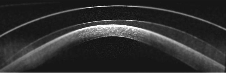
First, in this category it is good to realize that there is no 'static' single number representing scleral post-lens thickness (PLTL). Stephen Vincent et al found substantial regional variations in PLTL during scleral lens wear. The PLTL was measured across the central 5 mm at several time slots beteen 0 and 480 minutes after lens application using 12-radial-line scan optical coherence tomography imaging. A tilted optic zone was observed immediately after lens application, with the greatest PLTL asymmetry between nasal and temporal regions (156±22 μm more clearance temporally) and superior-nasal and infero-temporal regions (124±12 μm more clearance infero-temporally). The magnitude of lens settling observed in each region was associated with the initial PLTL. The superior-nasal PLTL furthest from the pupil center stabilized after 90 minutes compared with other regions, which stabilized after 4 hours. On average, after 8 hours of lens wear, the PLTL decreased by 29%, and PLTL asymmetries between opposing regions decreased by 30%. In summary: the PLTL was thickest temporally and thinnest nasally in healthy eyes fitted with rotationally symmetric scleral lenses, most likely because of regional differences in underlying scleral elevation, eyelid forces, and lens centration. PLTL asymmetries diminished with lens wear, and stabilization occurred more rapidly in regions with less corneal clearance immediately after lens application.
|
|
Scleral Lens Decentration vs. Apical Clearance |
|
Louise Kowalski et al investigated the effect of lens center thickness upon short-term horizontal and vertical scleral lens decentration as well as the association between both scleral topography and apical clearance with lens decentration. The mean scleral lens decentration was 0.55±0.19 mm temporally and 0.84±0.35 mm inferiorly, but this did not vary significantly with centre thickness. The mean nasal-temporal asymmetry in scleral elevation data was substantially greater (619±67 μm) compared to the vertical meridian (369±57 μm), and this variation in scleral topography along the horizontal meridian was associated with the magnitude of horizontal lens decentration. Greater initial central apical clearance was statistically significantly associated with more inferior lens decentration. In a previous study, Stephen Vincent and Michael Collins used scleral lens over-topography with a standard corneal topographer to quantify scleral contact lens decentration. Scleral lens decentration could be reliably quantified using tangential power maps with a standard (fixed) scale captured during over-topography without the need for customised instrumentation or image analysis software.
|
|
Measuring Inflammation in the Fluid Reservoir |
|
Ocular surface inflammation in scleral lens wear is longstanding concern and understanding the relationship between sclerals and the inflammatory state of the eye is essential, according to Maria Walker et al. This study is the first in its nature to compare the scleral lens fluid reservoir in healthy eyes and to compare them to basal tear samples after 8 hours and 4 days of scleral lens wear. In order to do so, fifteen normal habitual soft lens wearers were fitted with scleral lenses (with a diameter of 14.8 or 15.4mm). Tear components MMP-9 and MMP-10 were elevated in the fluid reservoir after several hours of scleral lens, suggesting potential clinical implications of scleral wear that deserves further investigation. The concentration of MMP-9 notably, and the diurnal variations in the fluid reservoir should be studied further, specifically in diseased eyes which may tend to produce more of this potentially damaging analyte. |
|
Fluid Reservoir Thickness and Corneal Edema |
|
There is debate concerning corneal oxygenation during scleral lens wear due to the potential additive hypoxic effect of a lens plus a fluid reservoir. The purpose of this study by Damien Fisher et al was to examine the effect of altering the fluid reservoir thickness on central corneal edema during short-term open-eye scleral lens wear and to compare these empirical measurements with predictive theoretical models. Scleral lens–induced central corneal edema increases with increasing reservoir thickness but plateaus at a thickness of around 600 μm, in agreement with recent theoretical modeling that incorporates factors related to corneal metabolism. Sandeep Dhallu et al found on this topic that a ≥125 Dk is advised for safe scleral lens daily wear. Scleral lens wear leads to an increase in corneal thickness regardless of lens-material oxygen permeability because of the fluid reservoir depth, they found. Mukesh Kumar et al found post–penetrating keratoplasty eyes to exhibit significant corneal edema after lens wear (2.99%), with greater swelling toward the graft-host junction inferiorly.
|
|
Influence of the Amount of Limbal Clearance |
|
This study by Debby Yeung et al is an objective and subjective evaluation of the clinical performance of scleral lenses with varying amounts of limbal clearance (LC) in keratoconus patients. Lenses with varying limbal clearances in 50 μm intervals were fitted and worn during a 2-week period. Participants reported greater comfort achieved with high LC. No difference in limbal and bulbar hyperemia was found, and there was similar perilimbal staining and negative corneal staining. In conclusion, the authors stated that the amount of LC may play an important role in subjective performance of scleral lenses but does not impact the degree of hyperemia in either the limbal or bulbar region. Although low LC might result in more compression-related changes to the ocular surface, high LC is associated with greater comfort and greater edematous changes. Limbal zone designs in scleral lenses might affect the integrity of the limbal epithelial tissue as a result of a combined hypoxic and compression-related etiology.
|
|
In line with the reported post-lens fluid reservoir issues mentioned, scleral lens fogging of the fluid behind the lens is an intrinsic scleral lens issue and unique to this modality. What can we do about it? |
|
Optical Density of Post-lens Fluid in Sclerals |
|
The purpose of this study by Turhan Akkaya et al was to determine the impact of optical density changes in post-lens fluid on clinical performance and to quantify these changes over time during miniscleral lens wear. The optical density changes behind the scleral lens were measured using Scheimpflug tomography at 30 minutes and at 1, 2, 3 and 4 hours. Compared with preoperative values, optical density significantly increased over time, while high-contrast visual acuity (VA) remained stable and low-contrast VA significantly decreased after two hours of lens wear. This seems in line with clinical findings from the scleral lens consulting room, as the study found that the optical density of post-lens fluid increases over time with miniscleral lens wear, with negative impact on low-contrast VA but not on high-contrast VA.
|
|
Factors Associated with Fogging |
|
A multicenter, cross-sectional study was conducted with an electronic survey that was distributed to scleral lens practitioners. Of the 248 survey respondents who indicated whether their patients had midday fogging, 26% reported midday fogging in this sample of established scleral lens wearers, according to Muriel Schornack et al. Midday fogging was not associated with age, sex, or race/ethnicity, and indications for scleral lens wear were not predictive of midday fogging. Neither specific lens designs (diameter or haptic design) nor care products were found to be associated with midday fogging. Patients who reported redness or irritation with scleral lens wear more commonly reported midday fogging. Data presented here suggest that this phenomenon may have a very complex origin. Although neither a particular lens-fitting philosophy nor a specific care regimen appear to reliably mitigate symptoms of fogging in general, it should be kept in mind that changes in characteristics of lens fit or care products may influence the development of fogging in individual patients. The association between redness or irritation with scleral lens fogging suggests that ocular surface inflammation may play a role in the accumulation of debris in the post-lens fluid reservoir. This also highlights the importance of timely and aggressive management of any signs of inflammation in scleral lens patients.
|
|
Midday Fogging with a New Scleral Lens Solution |
|
The purpose of this open-label study by Jennifer Swingle Fogt et al was to assess the safety of a scleral lens filling solution that closely approximates the ionic concentration and pH of human tears as well as to assess signs and symptoms of midday fogging with this formulation and with subjects' habitual sodium chloride solutions. The median Ocular Surface Disease Index score decreased from 27.1 when using habitual filling solution to 9.1 when using the test solution. Current Symptoms Survey findings with the test solution compared with habitual solution resulted in statistically significant decreases in burning/stinging, grittiness/foreign body sensation, dryness, blurry/fluctuating vision and overall pain/discomfort. Objective assessment of corneal staining and fogging revealed decreases that were not statistically significant in this small sample size. Further studies of the effectiveness of this solution in reducing midday fogging are warranted, as it looks promising at first glance, the authors state.
|
|
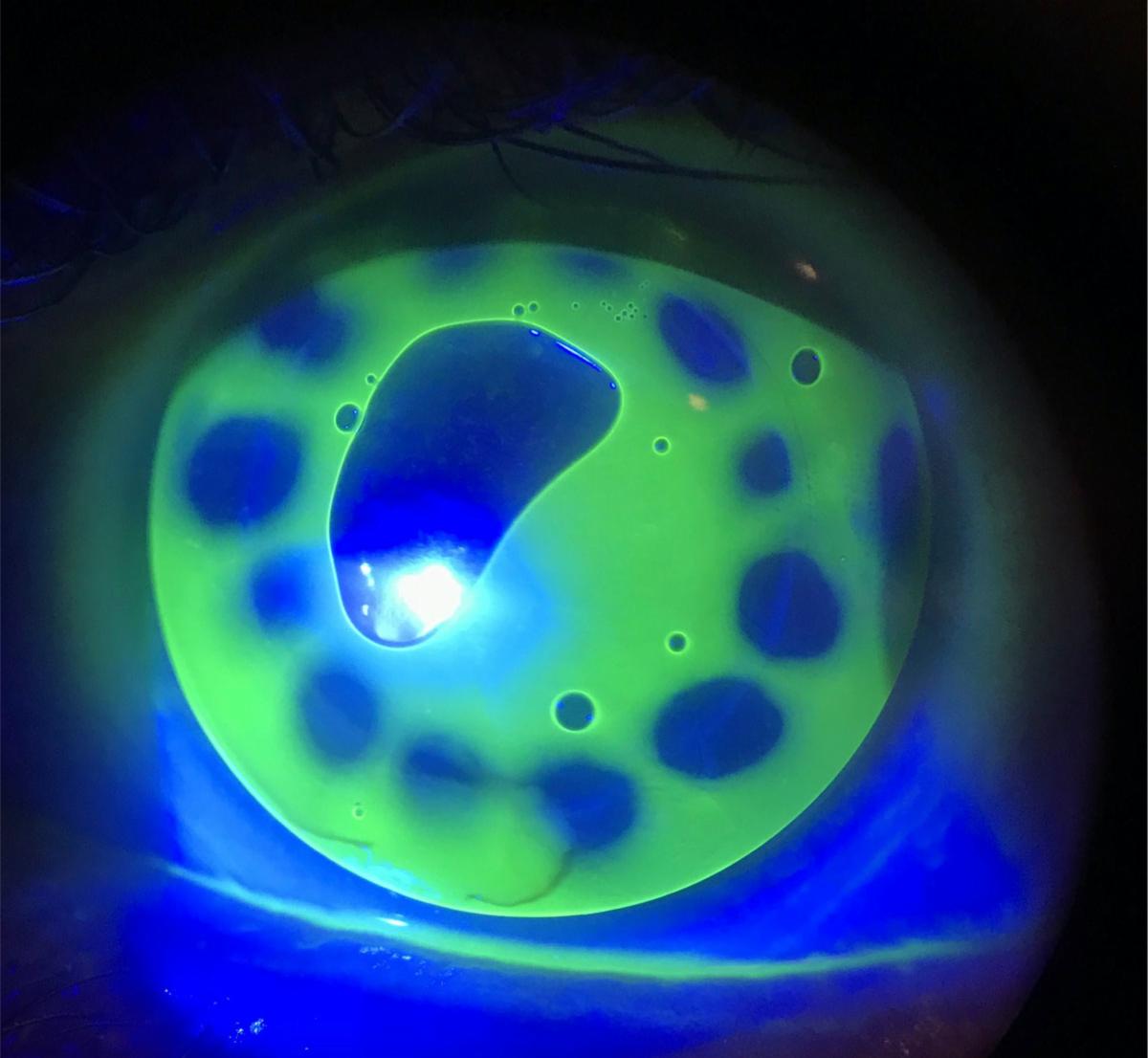
A fluorescein image of an intralimbal corneal (R)GP lens on a sunken corneal transplant. The steep central base curve causes a central bubble. Patients who desire the optics of a corneal GP with the comfort and protection of a soft lens beneath can consider a piggy back modality, which is often overlooked. This same corneal (R)GP lens was successfully piggybacked with a +7.00D silicone hydrogel soft lens. A patient’s comfort and tolerability of the lenses largely determines a successful piggyback fit. The soft lens and the GP should move independently of one another, and the (R)GP periphery should align nicely with the soft lens to avoid both adherence and excessive edge lift. From: Irregular Corneas Meet Their Match With GP Lenses, from Tiffany Andrzejewski and John Gelles in Review of Cornea & Contact Lenses.
|
|
Amsterdam, Netherlands
Copyright © 2020. All Rights Reserved
This newsletter is kindly supported by:
|
|
|
I-site is an educational newsletter that is distributed on a monthly basis and provides an update on rigid gas permeable-related topics (scientific research, case reports and other publications worldwide). I-site is objective and non-political. Disclosure: I-site's editor Eef van der Worp, optometrist PhD FAAO FBCLA FIACLE FSLS, receives educational grants from a number of industry partners but is not related to any specific company. Please contact us at:
|
|
|
|
|
|
|