I-site newsletter is a global educational newsletter FOR HEALTH CARE PROVIDERS launched in 2009, that monthly provides independent updates from the international literature on specialty rigid contact lenses such as corneal, orthokeratology and scleral lenses and related topics. View as Webpage
|
|
Keratoconus keeps intriguing—and also keeps surprising—us. The most downloaded and one of the most cited articles of the journal Contact Lens & Anterior Eye (BCLA journal) is 'Keratoconus: An Updated Review' by Santodomingo-Rubido et al. Maybe this should be considered in light of the fact that viewing keratoconus as a rare corneal disease with a prevalence previously estimated at 1:2000 is outdated. The latest new finding: in a population-based, prospective, monocentric cohort study in Germany, 10,419 subjects aged 40–80 years were included, with a 5-year follow-up. The study was published in Graefe's Archive for Clinical and Experimental Ophthalmology, which is not our lowest-ranked journal in the field when it comes to impact factor. Using Scheimpflug imaging, 75 eyes of 51 subjects were classified as having keratoconus. The prevalence for keratoconus in the German cohort, therefore, was 0.49% or 1:204 (with a 95% confidence interval of 0.36–0.64%). No gender predisposition was demonstrated. Logistic regression showed no association between keratoconus and age, sex, body mass index (BMI), thyroid hormone, smoking, diabetes, arterial hypertension, atopy, allergy, steroid use, sleep apnea, asthma, and depression in this sample. The investigators conclude that the prevalence of keratoconus in a mainly Caucasian population is approximately tenfold higher than previously reported in the literature using the latest technologies. As a profession and as an industry, it seems that we need to keep an eye on this disease.
|
|
To continue with the earlier remark that keratoconus is one of the most popular topics—even in non-specialty lens arenas such as Contact Lens & Anterior Eye, but also at the next NCC 2024 in the Netherlands (March 10-11), where keratoconus is one of the key (four) topics from the main stage—Contact Lens Update recently featured a full special on Corneal Ectatic Disorders.
|
|
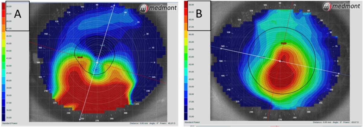
Daddi Fadel provides an overview on treatment options, including therapeutic contact lenses and surgical approaches. She explains that ectatic corneal disease comprises a group of disorders characterized by progressive thinning and protrusion of the cornea, leading to visual impairment, of which the most prevalent is keratoconus (KC). Among other things, she looks at differentiating corneal ectasias (such as KC from pellucid marginal degeneration [PMD] or keratoglobus). PMD is characterized by thinning and steepening of the inferior peripheral cornea, creating a characteristic “crab claw” or “kissing doves” topography pattern, while KC involves localized thinning and cone-shaped protrusion in the central cornea (Figure 1). PMD typically affects individuals in their fourth or fifth decade of life, while KC usually manifests during puberty. She also covers 'understanding inflammation in KC' and 'Individualized Management Approaches,' including an extensive flow chart on decision making in KC management, in which an elevation difference of ~350 microns on the ocular surface is considered a critical cut-off point in many cases. Erin Tomiyama in Optometry Times of May 30, 2023 also looks at KC management in all the different stages of the disease.
|
|
Current Perspectives in KC Management |
|
Melissa Barnett investigates the review article 'Current perspectives in the management of keratoconus with contact lenses' published by Lim et al in Eye. According to the reviewed cohort and cross-sectional studies, most patients with KC wearing contact lenses were fitted with corneal lenses. As for KC contact lens wear complications, these are associated with a variety of structural changes, such as decreased basal epithelial cell density, stromal keratocyte density, and endothelial cell density. KC was correlated with dry eye disease in a few studies and with an elevated prevalence of clinical signs, such as corneal staining and reduced tear break-up time, and a higher concentration of pro-inflammatory markers such as interleukins and metalloproteinases have been found in patients with KC. The contact lens type and material affect the risk of infectious keratitis. No evidence suggests that individuals with KC have an increased rate of infective keratitis compared to those wearing similar types of contact lenses.
|
|
Higher-Order Aberration & Vision |
|
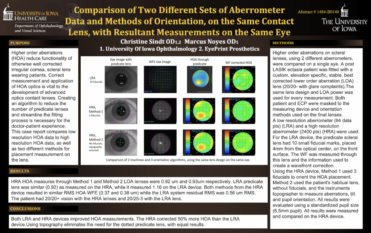
Christine Sindt visits the intriguing topic of higher-order aberrations (HOAs) in KC in a poster at the 2023 ARVO Annual Meeting, held in New Orleans in April. HOAs reduce the functionality of an otherwise well-corrected irregular cornea in scleral lens-wearing patients. Correct measurement and application of HOA optics is vital to the development of contact lenses with advanced optics. This case report compares two different sets of aberrometers—a low-resolution HOA device and a high-resolution device—on a single eye. A post-LASIK ectasia patient was fitted with a custom, elevation-specific, stable, best-corrected lower-order aberration lens (20/20- with glare complaints). All results were evaluated using a standardized pupil size (6.5mm pupil). They found that both the low-resolution aberrometer and the high-resolution aberrometer improved HOA measurements. In the clinical insight corner in Contact Lens Update, an advanced case of reclaiming vision and comfort in a KC patient is discussed by Karen Carrasquillo: 'Overcoming Higher Order Aberrations and Scleral Lens Suction' using a quadrant-specific scleral design with back-surface channels and haptic fenestrations.
|
|
Impact of Front-Surface Eccentricity |
|
To investigate the effect of front-surface eccentricity (FSE) of scleral lenses on corrected distance visual acuity (CDVA), contrast sensitivity (CS), and higher-order aberrations (HOAs) in keratoconic eyes, 15 keratoconic eyes were fitted with a scleral lens device of different eccentricity values (FSE0 = 0, FSE1 = 0.30, and FSE2 = 0.60). Baseline CDVA was 0.30 ± 0.29, which significantly improved to 0.10 ± 0.11, 0.05 ± 0.06, and 0.05 ± 0.08 logMAR with FSE0, FSE1, and FSE2, respectively. The post hoc showed no significant difference between eccentricities. Baseline CS was 1.19 ± 0.47, which improved to 1.54 ± 0.12, 1.59 ± 0.10, and 1.60 ± 0.11 with FSE0, FSE1, and FSE2, respectively, but post hoc again showed no significant difference between eccentricities. There was a statistically significant reduction in HOAs when compared with baseline. Comparison among eccentricities showed a significant difference only between FSE0 and FSE1 for coma and for spherical aberrations, for which FSE1 showed better performance in reducing HOAs compared with FSE0. Some key HOA root-mean-square values showed no significant difference between eccentricities, but FSE1 and FSE2 performed superior to FSE0. Practitioners should be aware of the optical impact of front-surface eccentricity as a distinct feature in scleral lens wear to improve visual performance. |
|
Epithelial Thickness A Key Element |
|
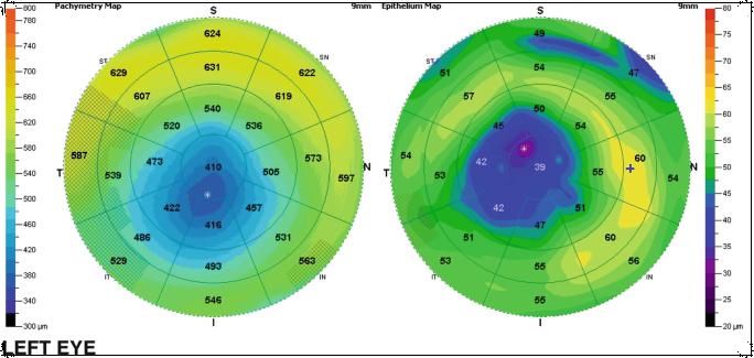
As mentioned in a post by Andrew Bowden on LinkedIn regarding early detection of keratoconus using epithelial thickness maps on OCT topography, epithelial thickness mapping can make a difference. This topic is covered in the recently published book 'Keratoconus' (Springer) by Márcia de Toledo and Bruno Gonçalves. They state that the epithelial thickness map is an auxiliary method for the diagnosis of keratoconus in eyes in which topography and tomography are suggestive but not conclusive. The two most widely available methods for obtaining epithelial maps are optical coherence tomography and high-frequency digital ultrasound. The main feature to understand is that the corneal epithelium can remodel itself to maintain the regularity of the epithelial surface despite the existence of irregularities in the stromal surface. In keratoconus patients, the epithelium tends to offset the underlying stromal ectasia tapering in the apex region of the cone by thickening around it. This behavior generates a pattern that resembles a “donut.” Lisa Stewart in Ophthalmology Times Europe also covers this topic in detail, pointing at the epithelial pliability. She makes several fierce statements: the epithelium thickens to fill any relative depressions, the epithelium thins over relative peaks within the stroma and the change in the epithelium is proportional to the change in the stroma.
|
|
The National Keratoconus Foundation held a webinar on "Risk Factors for Keratoconus" on May 9, 2023. A video recording is available on the NKCF website. Chantelle Mundy, clinical associate professor in the department of ophthalmology at The Ohio State University, seeks answers to questions such as 'Who gets keratoconus?' and 'Why?' Risk factors for keratoconus are described, including environmental triggers such as allergies and eye rubbing. The ethnic and genetic factors that put certain groups at a higher risk for the disease are addressed too as well as why certain family members are affected while other are not.
|
|
Amsterdam, Netherlands
Copyright © 2023. All Rights Reserved
This newsletter is kindly supported by
Bausch + Lomb Specialty Products
|
|
|
I-site is an educational newsletter that is distributed on a monthly basis and provides an update on rigid gas permeable-related topics (scientific research, case reports and other publications worldwide). I-site is objective and non-political. Disclosure: I-site's editor Eef van der Worp, optometrist PhD FAAO FBCLA FIACLE FSLS, receives educational grants from a number of industry partners but is not related to any specific company. |
|
|
|
|
|
|