I-site newsletter is a global educational newsletter FOR HEALTH CARE PROVIDERS launched in 2009, that monthly provides independent updates from the international literature on specialty rigid contact lenses such as corneal, orthokeratology and scleral lenses and related topics. View as Webpage
|
|
We fit more large-diameter lenses than small-diameter lenses in our practices, whether soft, hybrid or scleral. Yet, most of us have no access to a topographer that goes beyond the corneal borders. A new paper by Stefan Bandlitz and colleagues from Cologne (Germany) sheds some new light on the question: Can we predict, at least to some degree, the sagittal height (OC-SAG) of the ocular surface using Placido-based topographers? The answer is yes; quite literally, 'to some degree' we can. For the 8mm and 10mm corneal region, 94% and 89% of the variance of OC-SAG could be predicted using the central corneal radii alone. By including corneal eccentricity, this goes up to 95% and 92%, respectively. For 12mm, 14mm and 16mm regions, the authors found that when using central corneal radii alone, this was limited to 72%, 55% and 44%, respectively. Adding corneal eccentricity into the model increased the respective predictability to 84%, 71% and 60%. The best method to predict the OC-SAG is to use combined central radii, eccentricity and corneal diameter (86%, 78% and 65%). Interestingly though, the combination of just central radii and corneal diameter provided a slightly weaker prediction model than the combination of central radii and corneal eccentricity did. In contrast, corneal astigmatism was correlated only with corneal sagittal toricity and not with scleral toricity. In other words, scleral toricity and the axis of the flattest meridian seem to be independent from Placido-obtained corneal parameters and require additional measuring tools. But measurement of the SAG height in the corneoscleral region at a chord length equal to the overall lens diameter appears to provide an appropriate starting point when estimating the required contact lens sagittal height of a soft or scleral lens, the authors state.
Eef van der Worp
|
|
Conjunctival Vascular Density |
|
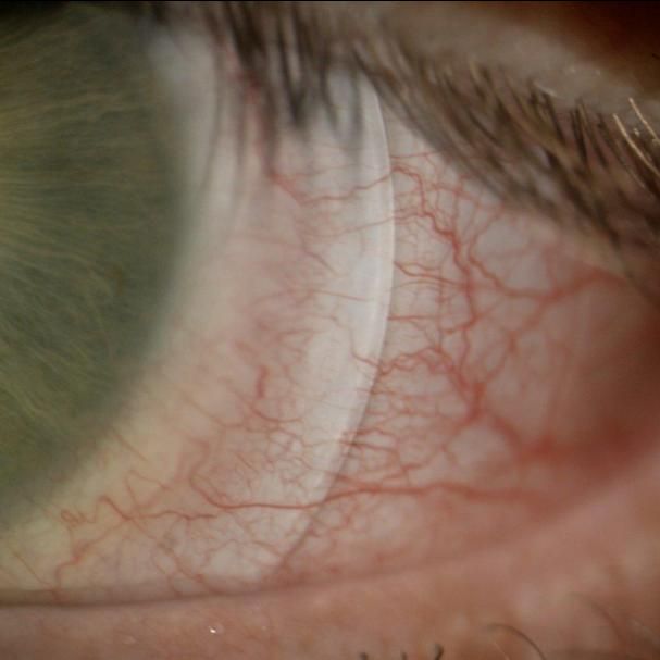
This study by Jennifer Jesus et al from Portugal investigated conjunctival vascular density using anterior segment optical coherence tomography angiography in scleral lens wearers. Patients who had keratoconus and were wearing scleral lenses (23) were included in the study. The conjunctival vascular density was significantly higher without a scleral lens than with a scleral lens in this group. A moderately negative correlation was found between indentation and vault of the scleral lens, and a positive tendency regarding the time of wearing scleral lenses and scleral indentation was observed. In conclusion, this method could be feasible to monitor conjunctival vasculature in scleral lens wearers. The results cautiously suggest that the repeated use of scleral lenses can cause vascular alterations in the conjunctiva, possibly due to a hidden hypoxia caused by prolonged limbal indentation, according to the authors. |
|
Endothelial Morphometric Changes |
|
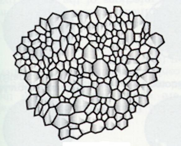
Amira Elagamy and Norah Al Omair from Egypt and Saudi Arabia looked at the correlation between long-term use of rigid corneal lenses and endothelial morphometric changes in keratoconus (KC) patients. The study included 40 KC eyes of non-lens-wearing patients (20 mild KC eyes and 20 moderate KC eyes) and 38 KC eyes with a long history of corneal rigid gas permeable lens wear (18 mild KC eyes and 20 moderate KC eyes). S pecialty KC lenses with a three-point-touch fit were used, and wearing duration in hours in mild KC eyes showed a significant moderate negative correlation with endothelial cell density (ECD) change and a moderate positive correlation with coefficient of variation of cell area (COV). In addition, wearing duration in years in moderate KC eyes showed a significant moderate negative correlation with the same variables (ECD and COV). Wearing duration in hours in moderate KC eyes, however, only showed a significant moderate negative correlation with percentage of hexagonal cells but not with ECD and COV. Significant corneal thinning in lens-wearing eyes was found in contrast to non-wearers. In conclusion, the study documented a significant correlation between long-term use of corneal specialty KC lenses with a three-point-touch fit and corneal endothelial morphometric changes. In addition, the current study confirmed significant corneal thinning in rigid corneal lens wearers compared to non-contact-lens-wearing KC patients.
|
|
Meibomian Gland Loss in Rigid Lens Wear |
|
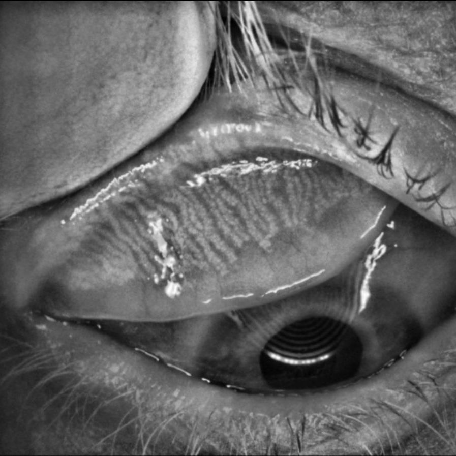
Ibrahim Inan Harbiyeli et al from Turkey looked at meibomian gland loss in rigid and in soft lens wearers. The prospective, multi-center, cross-sectional study included 44 soft lens wearers, 21 rigid corneal lens wearers, and 26 healthy non-wearers. The mean percentage of meibomian gland loss was 25.3% in soft, 34% in rigid and 18.4% in the control group. The OSDI (Ocular Surface Disease Index) scores in this study were correlated with mean meibomian gland loss in the lens-wearing groups. In a regression model including both lens groups, age and rigid lens material were predictive variables for mean meibomian gland loss. In conclusion, contact lens wear may cause meibomian gland dysfunction, and this effect may be more pronounced in rigid lens wearers. In soft lens wearers, the duration of lens use is an important variable associated with meibomian gland loss, and subjective symptoms could guide the prediction of meibomian gland loss in these cases.
|
|
Fluorescein & Lissamine Green |
|
Effect of Time on Grading
Corneal and Conjunctival Staining
|
|
We use these dyes all the time in our practices, but what is the effect of time on grading corneal fluorescein and conjunctival lissamine green staining in dry eye disease (DED)? This study found that the time required to reach the maximum grade of corneal fluorescein staining varied widely and was significantly longer in subjects with Sjögren's disease. This was not the case for conjunctival lissamine green staining. Early observation of corneal fluorescein staining can lead to under-grading, which may impact the diagnosis and assessment of treatment in DED. The results of this study underscore that the time of observation is an important factor in the assessment of corneal fluorescein staining, while the appearance of lissamine green staining is much less variable in the first 5 minutes following dye instillation. While these results apply only to the conditions in this study, the authors state that they hope this will provide a “wake-up” call for standardizing the use of fluorescein in the assessment of corneal staining in clinical practice as well as in clinical trials. |
|
Global Specialty Lens Symposium 2023 |
|
Call for Free Papers & Posters |
|
The Education Committee of the Global Specialty Lens Symposium invites the submission of abstracts for the presentation of Free Papers and Posters to be held January 18-21, 2023 at the Horseshoe Las Vegas, US (formerly Bally’s). The committee seeks topics including new and innovative concepts, clinical cases and research on all aspects of contact lenses (such as materials, designs, lens care, etc.) in addition to related topics such as corneal and ocular surface disease, diagnosis and treatment approaches and practice management. The submission window opens via the link below on July 1st - the deadline is October 3, 2022. |
|
Amsterdam, Netherlands
Copyright © 2022. All Rights Reserved
This newsletter is kindly supported by:
|
|
|
I-site is an educational newsletter that is distributed on a monthly basis and provides an update on rigid gas permeable-related topics (scientific research, case reports and other publications worldwide). I-site is objective and non-political. Disclosure: I-site's editor Eef van der Worp, optometrist PhD FAAO FBCLA FIACLE FSLS, receives educational grants from a number of industry partners but is not related to any specific company. |
|
|
|
|
|
|