I-site newsletter is a global newsletter, educational in nature and launched in 2009 that monthly provides independent updates from the international literature on specialty (R)GP lenses and related topics. |
|
A (W)hole New Perspective |
|
Sometimes, unexpectedly, new light is shed on apparently insoluble mysteries simply through smart and creative thinking. In the scleral lens field, it has been hypothesized for half a century that intraocular pressure (IOP) could be elevated during scleral lens wear. The main caveat, and why the discussions have been ongoing for such a long time, is that you cannot measure IOP on the cornea 'through the lens.' You can remove the lens, but that would alleviate the possible mechanism behind the cause of potential IOP increase. So any 'before' and 'after' lens wear analyses have their obvious limitations. An alternative method of measuring IOP through the eyelid, as previously reported on here, is a way to bypass that - but it is unclear whether the method itself of measuring through the eyelid is valid. The 'boys from Queensland' Samual Cheung, Michael Collins and Stephen Vincent came up with a really smart and simple alternative, published in Contact Lens & Anterior Eye. Why not put a hole in the lens? Through a central 4 mm fenestration, the IOP could be measured directly on the cornea. They found the IOP to be increased by up to 4 mm/Hg during lens wear, compared to immediately before and after lens wear. Still, there is room for speculation: the investigators hint that temporary compression of the ocular surface beneath the scleral lens may potentially be responsible for the IOP rise or that there is an alteration in aqueous outflow during scleral lens wear, which could artificially or directly elevate IOP values. That would be a whole different perspective. Eef van der Worp
|
|
Anterior Eye Surface Shape |
|
Scleral Shape in Emmetropes vs. High Myopes |
|
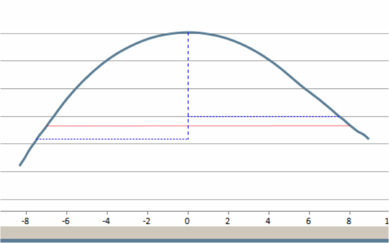
This newsletter frequently reports on both scleral shape and on myopia management. In the May edition of I-site newsletter, it was mentioned that 'it gets really interesting' when these two worlds collide; this item is a sequel to that. The group at Queensland University of Technology, Brisbane (AU) compared anterior scleral shape in emmetropes, low to moderate myopes, and high myopes with the Eye Surface Profiler instrument. In conclusion: high myopes exhibited a different anterior eye surface shape than emmetropes, having greater sagittal height in the nasal corneal periphery and anterior sclera. There was less nasal-temporal asymmetry of sagittal height and axial radius of curvature in high myopes than in emmetropes. Asymmetric growth of the eye associated with myopia development may be the underlying reason. These findings have implications for design of contact lenses, particularly soft and larger rigid lenses such as mini-sclerals.
|
|
Another hint on a potential link between scleral lenses and myopia is a study on the effects of accommodation and simulated convergence on anterior scleral shape by the same group. They found accommodation and simulated convergence to affect the nasal anterior scleral shape, with the greatest changes associated with convergence and being most evident in the more peripheral nasal scleral regions.
|
|
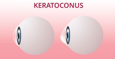
Despite advances in diagnostic tools, keratoconus is often diagnosed at a relatively late stage. Earlier detection of keratoconus would increase the overall clinical benefit of different therapeutic options. Elke Kreps and colleagues studied an impressive number of 399 patients (722 eyes) who had a mean age of 24.7±6.5 years, with an average maximal keratometry of 51±5.2 D for the better eye and 58.4±9.6 D for the worse eye. Upon diagnosis, 233 eyes (32.2%) and 51 eyes (7.1%) had a thinnest pachymetry of <450 and <400 μm, respectively. At 6-month follow-up, 58% of patients had been fitted with specialty contact lenses. During follow-up, 199 eyes (27.6%) underwent corneal crosslinking. Only one patient underwent corneal graft surgery of his worse eye due to contact lens intolerance and insufficient visual acuity.
|
|
In another study by the same research group from Antwerp (Belgium), the repeatability of an extensive number of relevant indices with the Pentacam HR in keratoconus detection was studied by Kreps et al. In subclinical keratoconus, repeatability did not differ significantly from controls (healthy volunteers), except for wavefront aberrations. In mild keratoconus, however, many parameters showed significantly worse repeatability compared to controls, and the repeatability of most parameters worsened in moderate disease. In particular, maximal keratometry and anterior astigmatism showed significantly worse repeatability in moderate keratoconus.
|
|
As Karen DeLoss writes in Contact Lenses Today of Sept 20, monitoring keratoconus and corneal progression can be a daunting experience. The mentioned research group from Belgium (Issarti et al) aimed at developing an objective severity scoring system for keratoconus for use in clinical practice. The resulting logistic index for keratoconus (Logik) provided consistent and progressing scoring that reflected keratoconus severity. The system provided an accurate classification of suspect versus normal keratoconus, with a sensitivity of 85.2% and a specificity of 70.0%.
|
|
Technology In Contact Lens Practice |
|
The anterior ocular surface is our workbench - no matter what lens we would like to fit. Two articles in Contact Lens Spectrum focus on this. Greg DeNaeyer in an excellent overview looks at scleral shape and how to measure it, versus its application for scleral lens fitting. A number of cases demonstrate how scleral shape measurement and assessment led to successful scleral lens design such as in post-RK corneal irregularities, scleral asymmetry and nasal pinguecula, and a custom design for a disciform scar. "We now know that most eyes that need scleral lenses have significant amounts of scleral asymmetry. This means that practitioners should be designing more customized lenses to achieve the best fit. This is particularly true for many patients who have corneal ectasias."
|
|
Another paper in the same Spectrum issue tries to cover corneal topography in depth, almost as a 'no-fee CE' topography course. It includes a 'top 10' list of how to take a good image (including regular callibration of the instrument, the use of the right capturing technique and use of the proper scales). As corneal topography is increasingly used to (directly) design corneal GP lenses, this means 'garbage in = garbage out" when it comes to lens manufacturing. This part is followed up by a very practical approach to analyzing a corneal topography map by Randy Kojima. For instance, when to consider a corneal GP versus a scleral lens? Lynette Johns brings it home by presenting a number of illustrative case reports.
|
|
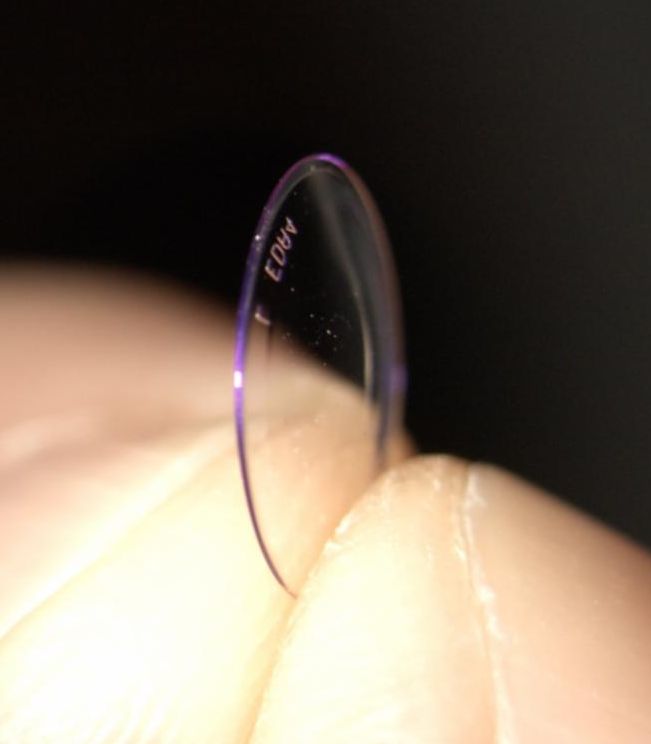
The pubquiz question that should accompany this image could be: 'is this lens for a cornea plana (an extremely flat cornea)? The answer: it is quite the opposite. It is a Rose-K2 Boston XO2 lens for a keratoconus patient that is everted. This case by Maaike Jansen from the Visser contact lens practice in Utrecht (the Netherlands) is about a 19-year-old female keratoconic patient who complained that the lens would 'pop out' regularly. The picture here shows the inside-out corneal GP lens. It is unclear why and how the patient, who was still in the adaptation phase, managed to evert the lens. The lens was replaced and the new lens worn successfully.
|
|
Thomas Arnold and Melissa Barnett host a new podcast, that provides 'a nexus for eye care practitioners all over the world' in their own words. Each month, a specialist from a different country will be featured for a lively discussion of the status of the eye care profession in their country. In this first episode the Australian pioneer of scleral lenses and a true trailblazer in scleral lens development Donald Ezekiel is interviewed. The podcast is educational in nature, but with a very personal touch and provides interesting personal details.
|
|
Amsterdam, Netherlands
Copyright © 2020. All Rights Reserved
This newsletter is kindly supported by:
|
|
|
I-site is an educational newsletter that is distributed on a monthly basis and provides an update on rigid gas permeable-related topics (scientific research, case reports and other publications worldwide). I-site is objective and non-political. Disclosure: I-site's editor Eef van der Worp, optometrist PhD FAAO FBCLA FIACLE FSLS, receives educational grants from a number of industry partners but is not related to any specific company. Please contact us at:
|
|
|
|
|
|
|