I-site newsletter is a global educational newsletter FOR HEALTH CARE PROVIDERS launched in 2009, that monthly provides independent updates from the international literature on specialty rigid contact lenses such as corneal, orthokeratology and scleral lenses and related topics. View as Webpage
|
|
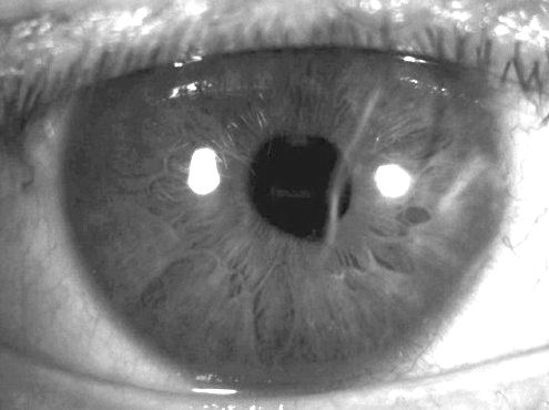
There are some questions you always wonder about in clinical practice, but you can't always get an answer. Here is one: in the discussion on Acanthamoeba infection & contact lens wear, we are encouraged not to use tap water. This is also true in gas permeable lens wearers. Our patients complain, saying that water from a tap simply rinses so nice. Is rinsing with a saline solution the same? Kelsey Steele and Karla Zadnik from The Ohio State University (US) investigated the rinsing options for rigid corneal lenses (RCLs) and assessed the efficacy of various saline solutions as an alternative method to tap water. The exiting fluid pressure of five commercially available saline solutions was measured using a venous pressure transducer system, and RCLs were cleaned with one of two commercially available cleaners and then rinsed with one of five saline solutions or with tap water. High-resolution imaging was performed after each rinse, and light transmission through the lens surface was determined by measuring the mean pixel count within one square millimeter. The mean pixels/mm2 and the rinsing method were correlated. High pixel counts represented more lens surface deposits and residue, and as fluid pressure of the rinsing methods increased, the mean pixels/mm2 decreased. These results imply that some products leave less residue compared to others on the lens surface - but tap water, with its high exiting fluid pressure, is the most effective option for removal of rigid lens cleaners from the lens surface. Despite these findings, tap water rinse is still discouraged because of its potential to result in corneal inflammation and infection; therefore, the development of alternative procedures and products is needed. In short, we need more rinsing power! At least we have clarity on this now—it 'cleans up' nicely what we knew about the subject.
|
|
Xiao-Di Yang and Yong Lyu from Zhengzhou University in China report that children are more likely than adults to develop amblyopia after ocular trauma; thus, timely visual rehabilitation after ocular trauma in children is important. Most open-globe injuries are accompanied by corneal laceration, and the corneal opacity and corneal irregular astigmatism caused by corneal scarring are important factors that affect vision. In the past few decades, visual rehabilitation of corneal scarring after corneal laceration has been managed by procedures such as corneal transplantation and by (rigid) contact lens correction. That said, few studies have reported the use of RCLs for refractive correction after corneal laceration in children younger than 12 years. The hypothesis of this study was that wearing RCLs after corneal trauma in children results in better visual outcomes compared to wearing spectacles. The findings show that the best-corrected visual acuity statistically significantly improved with RCLs, but six of the 15 children abandoned wearing RCLs because of discomfort and lens rejection (n=3), lens loss and inability to replace broken lenses because of travel distances and epidemics (n=2), and cost (n=1). Despite considerations of lens application and initial wearing comfort, RCLs can be a beneficial and safe tool for postoperative visual rehabilitation in children with open ocular trauma, the study concludes.
|
|
Post-Lens Tear Film Thickness in RCLs |
|
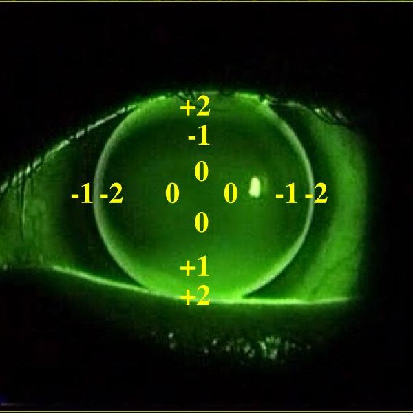
Another one of these 'why don't we know this' questions (see column above) is: What is the correlation between base curve radius (BC) changes and the post-lens tear film thickness in microns RCLs? The button below links to one of the few studies in the field on this topic from Poland. The authors measured the post-lens tear film thickness with an anterior segment spectral optical coherence tomography (AS-SOCT) device for assessing the lens-to-cornea fit of RCLs. Fourteen patients (26 eyes) were enrolled, and the initial BC of each lens was determined based on keratometry readings. The fluorescein pattern and tomograms were evaluated, starting with an alignment fit and, subsequently, with BC reductions of 0.1mm, 0.2mm and 0.3mm. Repeatability of the measurements was 18%. BC reductions of 0.1mm, 0.2mm, and 0.3mm resulted in average apical clearances of 12.38 (SD=9.91), 28.79 (SD=15.39), and 33.25 (SD=10.60), respectively. In conclusion: BC steepening of 0.1mm or more led to measurable changes in lens-to-cornea fits. Interstingly: although AS-SOCT represents a new method of assessing lens-to-cornea fit, apical clearance detection with current commercial technology showed lower sensitivity than the fluorescein pattern assessment. |
|
Getting in Shape on the Corneo-Scleral Junction |
|
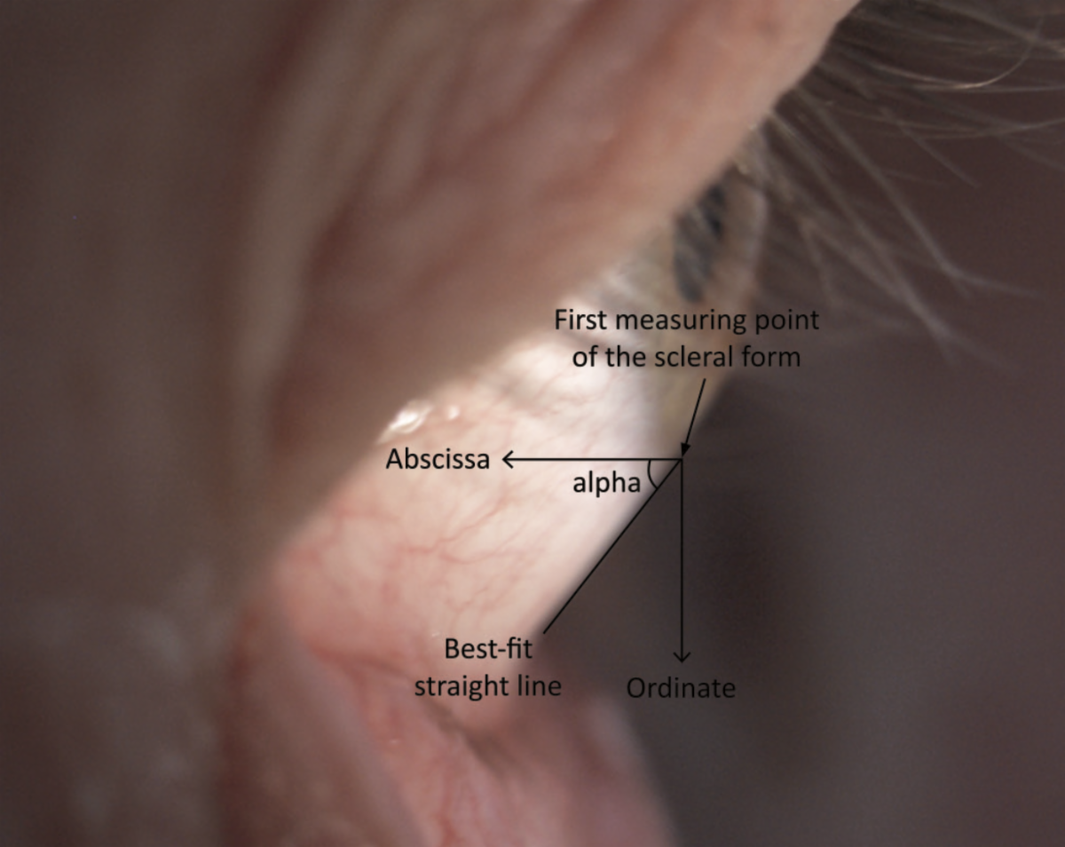
The aim of this study from Germany published in Optometry & Contact Lenses (OCL) was to present a theoretical modelling method to improve the fitting of scleral lenses by means of a novel determination of the anterior scleral shape. The authors state that the movement of scleral lenses and soft lenses is affected by the scleral shape around the limbus, and thus it is important to take a closer look at the edge of the sclera. An area of 4mm in diameter around the limbus is considered a suitable area for shape fitting, as shown in 44 profile photographs. Based on the 44 profile photos, 85.8% of the corneo-scleral profiles (CSPs) were straight, 11.1% were convex and 3.1% were concave parabolic shapes. The probability of determining a parabolic CSP section is about 18.5%. Conclusion: a specification of the CSPs using this method (the scleral angle alpha) provides significantly more information about the scleral shape. Furthermore, it simplifies the determination of the geometry of large-diameter contact lenses. Photo: Manfred Bufler - OCL
|
|
Monitoring Patients Corneal Health |
|
Brooke Messer in 'orthokeratology in practice' from Contact Lens Spectrum walks us through practical tips and monitoring patients’ corneal health with ortho-k. Corneal staining and other findings can show up for various reasons during the ortho-k process. When we see, for instance, epithelial disruption on the central cornea after lens wear, the sagittal depth is likely too shallow and the lens is bearing on the corneal apex, Messer writes. Corneal staining can also occur when the lens is too deep or too tight in the peripheral curves, and a tight ortho-k lens will trap stagnant tears or possibly soaking solution under the optical zone and create a toxic response in the corneal epithelium. Corneal impression rings may also be noted during ortho-k lens wear, and patients with these often complain of difficulty or pain with lens removal in the morning. Understanding why we see certain corneal changes throughout treatment can improve our ability to assess lens fits on-eye and promote long-term corneal health in our ortho-k lens wearers, she continues.
|
|
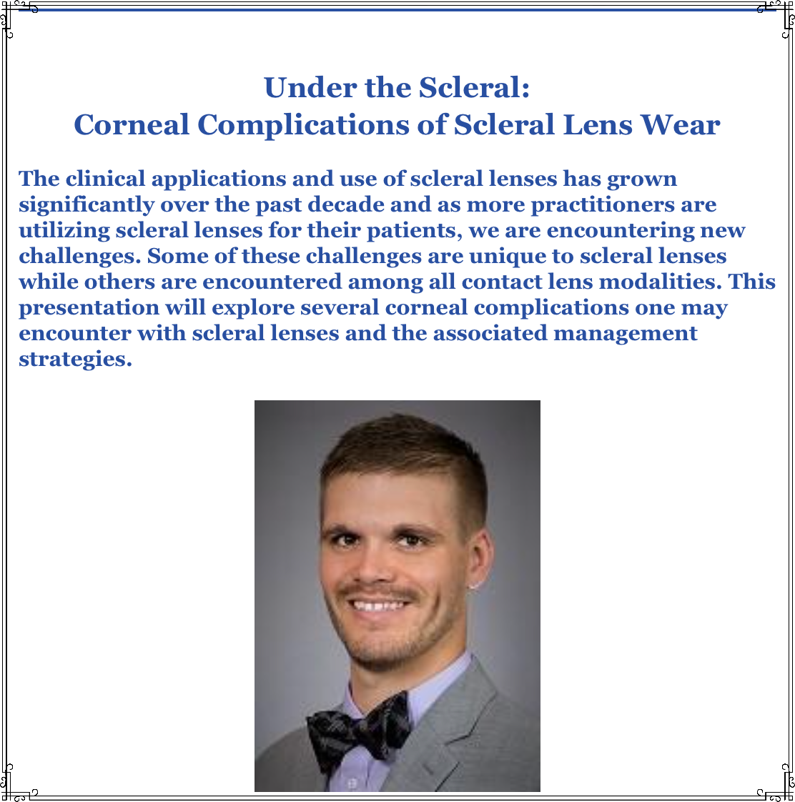
The clinical applications and use of scleral lenses have grown significantly over the past decade, and as more practitioners are utilizing scleral lenses for their patients, we are encountering new challenges. Some of these challenges are unique to scleral lenses, while others are encountered among all contact lens modalities. This webinar by Trevor Fosso from Minnesota (US), which is hosted by the Scleral Lens Education Society, explores several corneal complications one may encounter with scleral lenses and the associated management strategies. |
|
Amsterdam, Netherlands
Copyright © 2023. All Rights Reserved
This newsletter is kindly supported by
Bausch + Lomb Specialty Products
|
|
|
I-site is an educational newsletter that is distributed on a monthly basis and provides an update on rigid gas permeable-related topics (scientific research, case reports and other publications worldwide). I-site is objective and non-political. Disclosure: I-site's editor Eef van der Worp, optometrist PhD FAAO FBCLA FIACLE FSLS, receives educational grants from a number of industry partners but is not related to any specific company. |
|
|
|
|
|
|