Teaching Tuesday

Neointimal Hyperplasia and Restenosis

This Teaching Tuesday focuses on neointimal hyperplasia and restenosis.


Neointimal hyperplasia is the proliferation and migration of cells in the tunica intima, which causes a thickening of the arterial wall. This is a repair process that occurs in response to trauma to the vessel; the new cells are analogous to "scar tissue." 


Initially, this process is beneficial because blood flow travels over a layer of smooth cells instead of the stent, reducing the risk of clotting. However, as new layers are added over time, the diameter of the lumen can decrease significantly. Neointimal hyperplasia is the most common cause of restenosis after arterial intervention.


The demonstrative case below showcases neointimal hyperplasia within a stent post-endarterectomy.

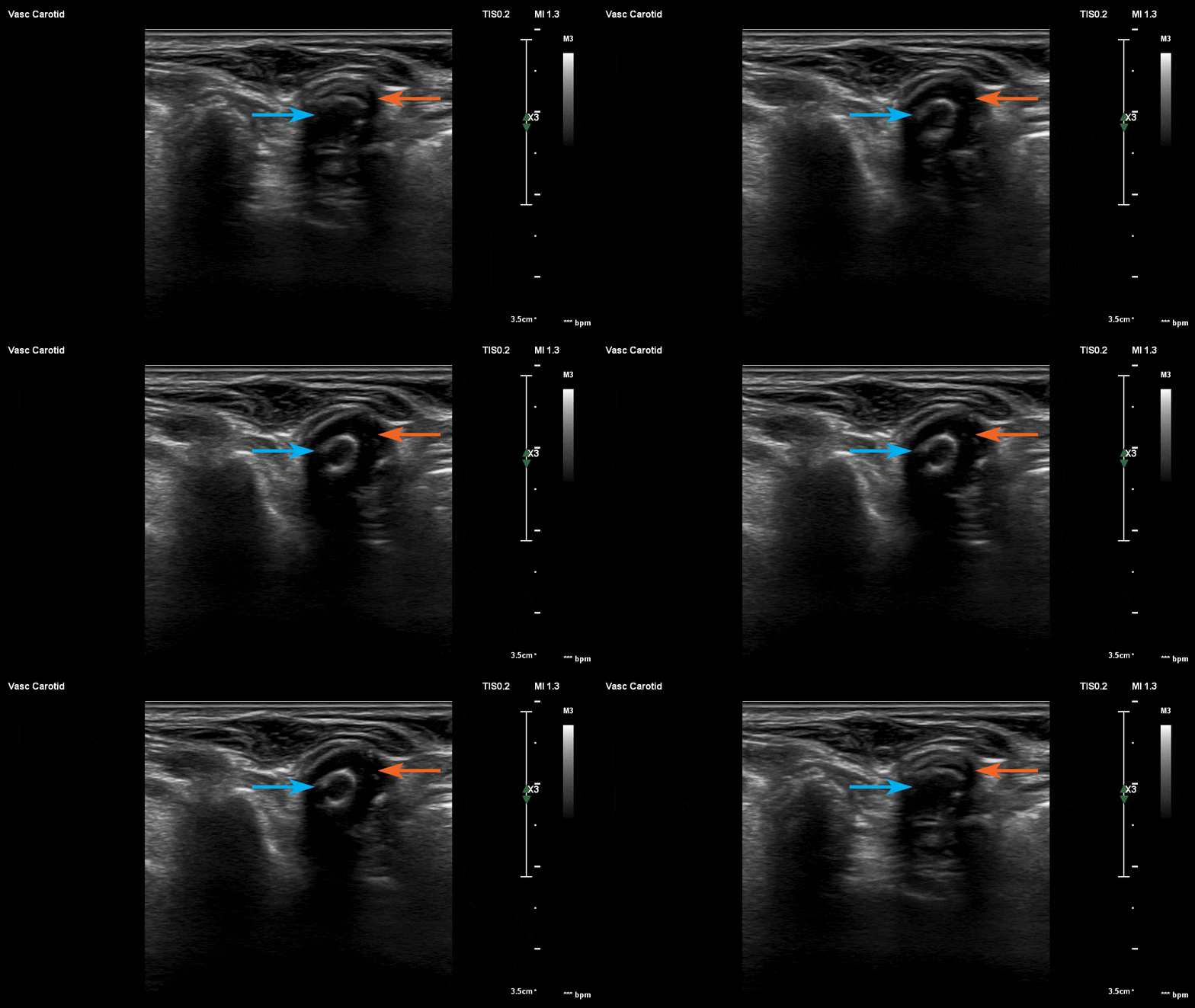
B-mode of the internal carotid artery in transverse view. Note the endarterectomy site (orange) and the stent (blue) within it.

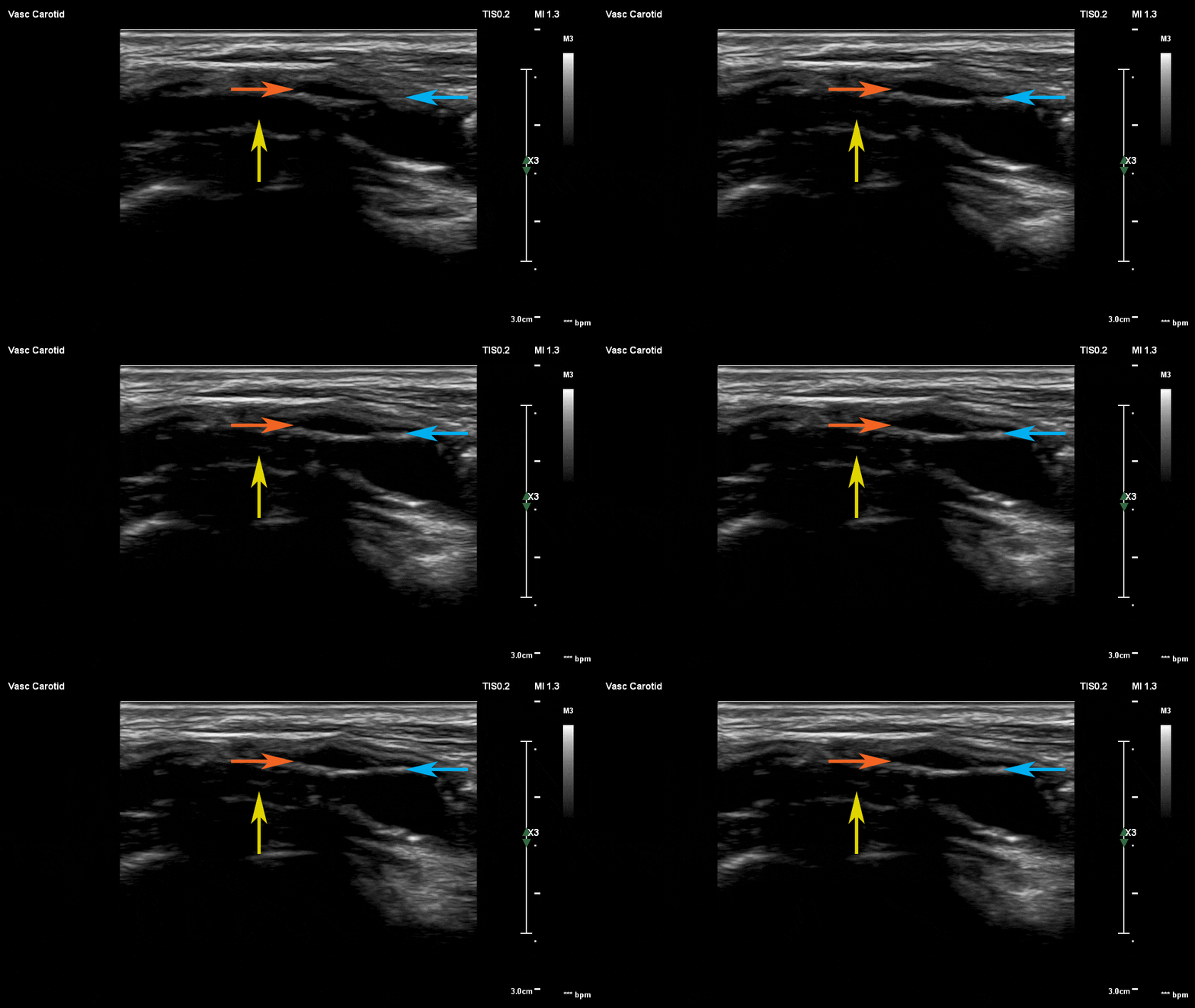
B-mode of the internal carotid artery in longitudinal view. Comparing with the previous image, note the endarterectomy site (orange) and the stent (blue) within it. Also, note the scar tissue within the stent caused by neointimal hyperplasia (yellow).  

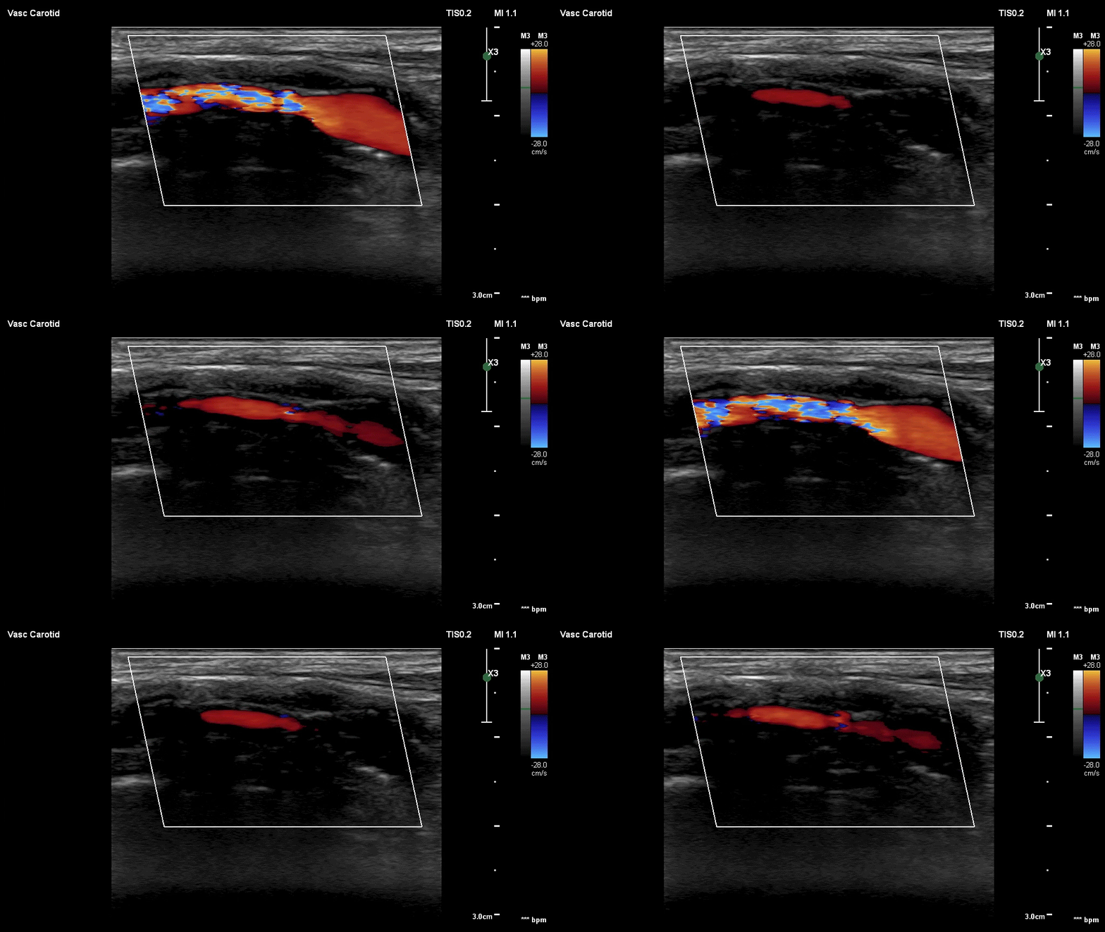
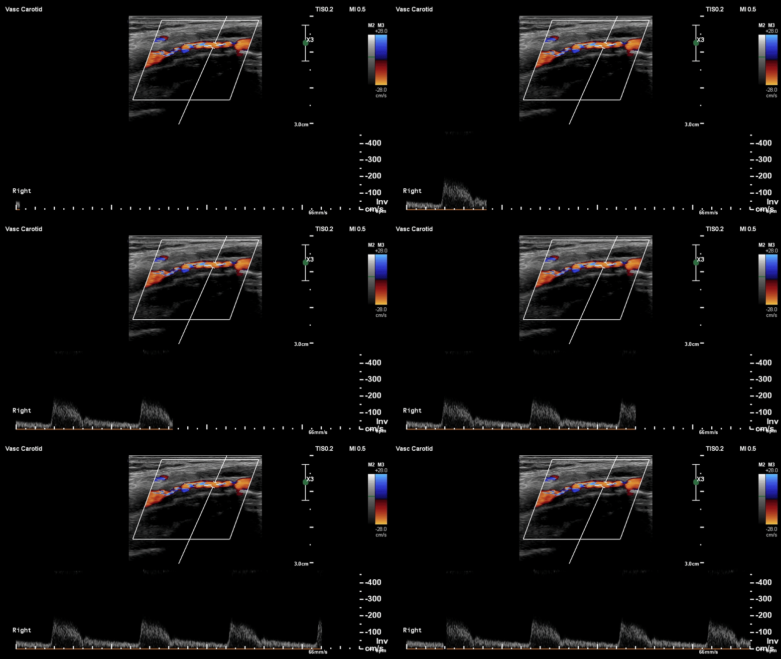
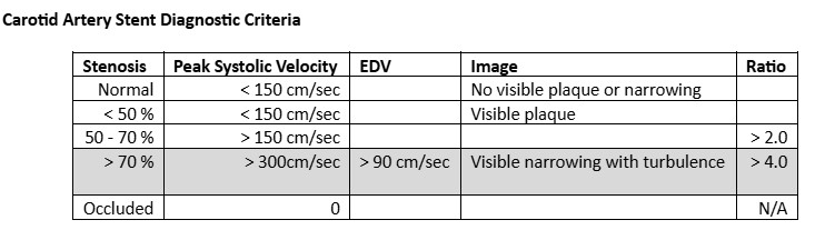
Color Doppler of the internal carotid artery in longitudinal view. Note the color aliasing. Due to Bernoulli’s principle, there is increased blood flow velocity through a stenotic segment. Color aliasing, which occurs due to increased velocity, is an indicator of restenosis in the stent.

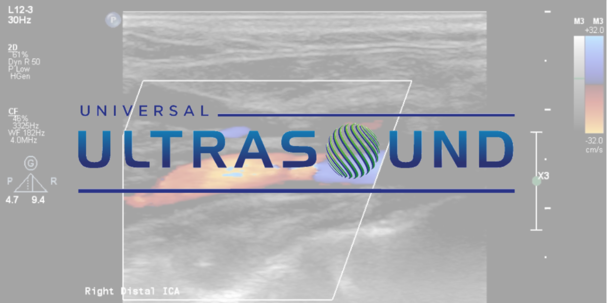
Finally, a combination of color Doppler and pulsed-wave Doppler of the internal carotid artery in longitudinal view. This combination allows you to see a real-time feed of the waveform as you insonate the length of the artery, which ensures that you do not miss any increased velocities. Note the increased peak velocity in this segment.

Purchase Course Now!

Would you like to learn more regarding neointimal hyperplasia as well as other physiology and pathophysiology that can be visualized via ultrasound? Join Julie Cardoso and team as they tackle live patient cases in our peripheral arterial course!

Facebook  Twitter  Instagram