|
Background
The brown recluse spider (Loxosceles reclusa) is one of two medically significant spiders in North America along with the black widow. While several Loxosceles species are present in the United States, L. reclusa has the widest distribution, spanning 16 states from southeastern Nebraska to southwest Ohio and south through Texas to northern Georgia. In nature, these spiders inhabit woodpiles, tree bark, and rocky debris; indoors they are typically found in garages, basements, and other undisturbed areas. Their webs are asymmetrical and lined with dense, white silk. Brown recluses are nocturnal hunters capable of surviving up to six months without food or water. Although generally non-aggressive, they bite when provoked, with females exhibiting greater defensive behavior. Most envenomations occur between spring and autumn, often in the morning (Repplinger, D.J. & Hahn, I., 2019).
Identification
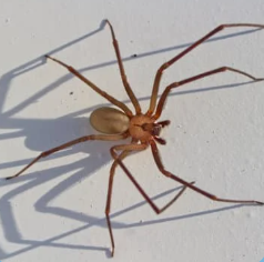
The brown recluse is also known as the “violin” or “fiddleback” spider, due to the dark violin-shaped marking on the dorsal cephalothorax. However, this feature is variable and not diagnostic, as similar markings occur in other species (e.g., cellar spiders, pirate spiders). A more reliable identifying feature is the unique eye pattern: six eyes arranged in three dyads in a semicircle, unlike the typical eight seen in most spiders (Repplinger, D.J. & Hahn, I., 2019).
Pathophysiology and Clinical Presentation
Brown recluse venom is cytotoxic, composed primarily of sphingomyelinase D and hyaluronidase. Sphingomyelinase D induces dermonecrosis, hemolysis, and serotonin release from platelets, while also activating inflammatory cascades involving prostaglandins, leukotrienes, neutrophils, and thromboxane. Hyaluronidase facilitates venom spread but does not directly cause tissue injury (White, J., 2017). The ratio of venom to spreading factor influences lesion severity. Clinically, most bites result in localized erythema, pruritus, and mild induration that resolve within weeks. Larger inocula can produce blistering, ulceration, and necrosis with eschar formation, often requiring 2–4 months for healing. Rarely, systemic loxoscelism develops 24–72 hours post-bite, presenting with chills, malaise, vomiting, arthralgias, jaundice, hemolytic anemia, and/or acute renal failure, independent of cutaneous severity. The incidence varies by region and Loxosceles species involved.
Treatment
Loxosceles envenomations are variable, and no U.S. approved antivenom is available (Swanson, D. L. & Vetter, R. S., 2005). Laboratory testing cannot confirm envenomation, though findings such as hemolysis and anemia may support diagnosis. Management is supportive, focusing on local wound care, prevention of secondary infection, symptomatic relief, and the treatment of systemic complications.
|