|
|
Quarterly Update
Arkansas State
Veterinarian's Office
|
|
|
Arkansas Department of Agriculture
Livestock and Poultry Division
|
|
Spring 2021 (Volume 10)
Randolph Chick, DVM (479) 200-2551- cell
|
|
Rabbit Hemorrhagic Disease (RHDV-2) has been confirmed in a domestic rabbit in northeast Arkansas. Rabbits can become infected through direct contact or exposure to an infected rabbit’s urine, feces, and blood. The virus can survive for months in the environment. People can spread the virus by carrying it on their shoes or clothing, contaminated feed/hay, using uncleaned cages and water bowls, or petting and carrying infected rabbits. Related links:
Click here to view the ARBA Rabbit Hemorrhagic Disease Policy.
|
|
Veterinary Medical Loan Repayment Program (VMLRP) – Final Date is April 16.
USDA’s VMLRP helps qualified veterinarians offset a portion of education debt in veterinary medicine in return for their services. The National Institute of Food and Agriculture (NIFA) will enter into educational loan repayment agreements with veterinarians who agree to provide their services in veterinarian shortage situations. Veterinarians must commit to providing at least three years of veterinary services in a designated veterinarian shortage area. NIFA may repay up to $25,000 of your student loan debt per year. Loan repayment benefits are limited to payments of the principal and interest, on government and commercial loans, received for study at an accredited college of veterinary medicine. Review the eligibility requirements to determine if you are eligible.
|
|
Livestock Exhibitions – It’s that Time Again!
Animals being shown within the state of Arkansas must be accompanied by a Certificate of Veterinary Inspection (CVI or health papers). Health certifications must be dated for use within the borders of Arkansas for 90 days from issuance - be sure to check with individual shows for differing requirements. Testing results for diseases of concern are to be noted on the CVI and should be repeated as required. A Category II Accredited, Arkansas licensed veterinarian needs to issue health certifications:
- Personally inspect animals and sign forms (i.e., test charts, ICVIs, other health certificates);
- Issue complete and accurate forms (i.e., all official IDs should be recorded);
- Indicate and verify work done by other accredited veterinarians (i.e. testing results);
- Immediately report incidence of reportable disease;
- Maintain biosecurity in conducting of medical and regulatory functions;
- Know current regulations;
- Have contact information on-hand for state and federal veterinarians responsible for their state (including the federal area veterinarian in charge and the state veterinarian); and
-
Please see 9 CFR Parts 106, 161, and 162 for the full regulations concerning the National Veterinary Accreditation Program (NVAP) and duties of an accredited veterinarian. USDA APHIS | A: 9 CFR PARTS 160, 161, and 162
|
|
As an Extension livestock veterinarian, I have the honor of wearing many hats. I train county Extension agents how to help livestock producers effectively and efficiently monitor and manage animal health on the farm. I also coordinate the Arkansas Beef Quality Assurance Program, which educates producers on best practices for beef cattle, and the Arkansas 4-H Veterinary Science Program that promotes veterinary careers to youth. For the last two years I have also been the attending veterinarian for the Arkansas State Fair, through an agreement between the University of Arkansas System Division of Agriculture and the Arkansas State Fair Board. The role of State Fair Veterinarian comes with much responsibility and requires keen eyes and strong knees.
As with all animal exhibits, there is an initial chaos when animals are checked in and escorted to their temporary enclosures. Inspectors for the Arkansas Department of Agriculture Livestock and Poultry Division have served as essential sentinels, posted at the livestock entrance to check animal health papers and guide exhibitors to their destination. Before the animals reach their reserved space at the barn, they must pass a health check through the attending veterinarian. With hundreds of animals to check in, the health check must be quick, yet thorough. Achieving efficient and detailed health checks mean every exam is the same, no matter which species is coming through. I have been asked to describe what I am assessing during a health check and what happens if I find something that disqualifies an exhibit animal. The easy answer is that I am looking for anything that could be infectious to people or to other animals.
The exam starts with the head. I look in the ears for any evidence of mites or infection, the eyes and nose for discharge that could signal a respiratory infection, and the mouth for any sores that could indicate a viral disease. Next, I check the skin of the entire body for crusty lesions that could be ringworm or masses that could be warts, and the feet for sores that could be infectious foot rot. This is done by looking and feeling at the same time. Finally, I visually inspect the area around the anus for signs of diarrhea. If anything is found, a closer look is given, and the exhibitor is asked questions. If what is found is determined as potentially infectious, the exhibitor is asked to either take the animal back home or place the animal in a pen arranged in the quarantine barn. I always make certain to explain the reason for disqualification to the exhibitor and give advice on how to prevent the problem in the future.
For sheep and goats, ringworm is the usual disqualifier. For cattle, warts are the most common disqualifier. This past year we also started examining rabbits upon entry for any signs of Rabbit Hemorrhagic Disease such as jaundice, diarrhea, or bleeding. After initial inspections, I monitor the animal facilities daily for anything unusual. If a health-related problem arises I am alerted by the livestock manager. The goal, as the attending veterinarian, is to keep infectious diseases away from the property. One infected animal can cause a massive spreading event. For several youth, and their families, their livelihoods depend on the health of their show animals. Although it breaks my heart to turn away an exhibit animal, I know I am doing so to protect all other animals.
|
|
Left to right: Wes Ward, AR Secretary of Agriculture; Dr. Heidi Ward, Asst. Professor of Animal Science and Veterinarian; and Warren Carter, Executive Vice President of Farm Bureau |
|
|
Exhibition/Show Season Biosecurity – Dr. John Nilz, ADA - Livestock and Poultry Division
Biosecurity is a set of practices that prevent the introduction and spread of diseases. Application of biosecurity is easier on isolated premises. Traveling and commingling during show circuits presents a more difficult challenge. This article addresses specific ideas and links to more information concerning the protection of show animals. Sections include material that can be applied to phases (Before, During, and After) of a show.
Before Show – Have your animals examined by a veterinarian within the required timeframe established by the state and show officials. After initial examination, continue to monitor the animals. Call your veterinarian if any signs develop during any of the phases. A suggested list includes but is not limited to the following:
- Any unusual changes in sounds such as coughing, sneezing, and/or wheezing.
- Any unusual discharges from eyes, ears, nose, mouth, skin, urinary tract, and the rectum.
- Any unusual changes in coloration of skin, urine, or stool.
- MOST importantly – BLISTERS anywhere on the body. This is a common feature of reportable and foreign animal diseases.
Clean and disinfect prior to departing for a show. Consult your veterinarian to find the appropriate products to use on ALL equipment and your animals. Place a high priority on maintaining a closed herd during the show season.
During Show – avoid direct contact with other animals and people. Exhibition management should provide a schedule for wash racks and orderly movement to tie-outs (if employed for your species). Do not share equipment of any kind. Examples include dressage equipment, feed, tubs, bedding, or tack.
After Show – diseases have an incubation period, so they may not show up immediately. Isolate your show animals from the main group for a period based on your attending veterinarian’s recommendations. If showing market animals, consider sending to finish or slaughter, and not returning them to the farm. Clean and disinfect all items prior to return.
Links:
|
|
Equine Infectious Anemia (EIA) – R. J. Chick, DVM, ADA-LPD, March 30, 2021
Around 35,000 Arkansas horses are tested annually for Equine Infectious Anemia (EIA), out of a reported population of around 160,000 in the state. Many potential reservoirs for the EIA virus are not being evaluated for the EIA threat on an annual basis, as called for in the Arkansas Code Annotated - A.C.A. § 2-40-804…(a) (1), “All equidae domiciled within the State of Arkansas shall be subjected to an official equine infectious anemia test every twelve (12) months. (2) An equidae is domiciled within the state when the equidae has been pastured, stabled, housed, or kept in any fashion in the state more than thirty (30) consecutive or unconsecutive days. (3) Written proof of a negative current official equine infectious anemia test shall be made available in the form of negative results from an approved laboratory upon request made by an authorized representative of the Arkansas Livestock and Poultry Commission….”
USDA reports for the year 2019 noted 89 EIA positive horses on 38 premises for all the USA. Arkansas had two detected that year; both animals were euthanized and buried on their pasture plot. Rescue animals coming from neighboring states are especially important to test prior to arrival in Arkansas and need to be monitored for any delayed development of the disease. Missouri recently detected EIA in two groups of horses; one had been on pasture for 4 years and had not been tested before addition to the herd, another location had a positive mare that had been purchased and tested 3 years prior to detection. Approximately 35 horses will be quarantined on the two Missouri premises for months of follow-up testing after the removal of the EIA reactors (to enable quarantine release).
|
|
Mycoplasmosis in Poultry – (Chronic Respiratory Disease, Infectious Sinusitis), M. El-Gazzar, DVM, MAM, PhD, DACPV, Department of Veterinary Diagnostics and Production Animal Medicine, College of Veterinary Medicine, Iowa State University - modified May 2020
Mycoplasma gallisepticum causes respiratory infections in chickens, turkeys, and other avian species. Morbidity is typically high and mortality low in affected flocks, and signs are generally more severe in turkeys. Real-time PCR is becoming the most common test used for diagnosis. Antibiotics may reduce clinical signs and transmission through eggs, but they do not eliminate infection. Control is achieved by good biosecurity and sourcing stock from M gallisepticum-free breeder flocks. M gallisepticum is commonly involved in the polymicrobial "chronic respiratory disease" in broiler chickens, leading to increased condemnations in the processing plant. In layers and breeders, it is usually subclinical, but causes a reduction in the number of eggs laid per hen over the production cycle. Turkeys are more susceptible to M gallisepticum, frequently resulting in swollen infraorbital sinuses and is thus called "infectious sinusitis." These diseases affect chickens and turkeys worldwide, causing the most significant economic losses in large commercial operations, and are commonly seen in noncommercial flocks. Infection also occurs in pheasants, chukar partridges, peafowl, pigeons, quail, ducks, geese, and psittacine birds. Songbirds are generally resistant, although there is a widespread outbreak of M gallisepticum causing conjunctivitis and mortality in wild house finches (and some similar species) in North America. M gallisepticum is the most pathogenic avian mycoplasma; however, considerable strain variability is manifest in a range of host susceptibility, virulence, clinical presentation, and immunologic response….
M gallisepticum is transmitted vertically within some eggs (transovarian) from infected breeders to progeny, and horizontally via infectious aerosols and through contamination of feed, water, and the environment, and by human activity on fomites (shoes, equipment, etc). Infection may be latent in some birds for days to months, but when birds are stressed horizontal transmission may occur rapidly via aerosols and the respiratory route, after which infection and clinical disease spread through the flock. Once individuals or flocks are infected, they remain infected for life and act as carriers or reservoir for infection. Flock-to-flock transmission occurs readily by direct or indirect contact from the movement of birds, people, or fomites from infected to susceptible flocks.
Some potential reservoirs of M gallisepticum in the USA are noncommercial (backyard) flocks, multiple-age layer flocks, and some free-ranging songbird species. Good management and biosecurity practices are necessary to ensure that M gallisepticum infections are not introduced to commercial poultry from these and other sources… once infected, birds may remain carriers for life. There is a marked interaction (polymicrobial disease) between respiratory viruses (Escherichia coli) and M gallisepticum in the pathogenesis and severity of chronic respiratory disease…. morbidity is high and mortality low in uncomplicated cases. Nasal discharge and conjunctivitis with frothiness about the eyes may be present. The disease is generally more severe in turkeys than in chickens and swelling of the infraorbital sinuses is common. Diagnosis is increasingly based on real-time PCR. History, clinical signs, and typical gross lesions may be suggestive of M gallisepticum infection. Serology by agglutination and ELISA methods are commonly used for surveillance. Hemagglutination-inhibition is used as a confirmatory test, because nonspecific false agglutination reactions may occur, especially after injection of inactivated oil-emulsion vaccines or infection with M synoviae. Mycoplasma isolates must be identified by species, because birds may also be infected with nonpathogenic mycoplasmas. E coli infection, Newcastle disease, avian influenza, and other respiratory diseases (i.e., infectious bronchitis in chickens) should be considered in the differential diagnosis, and can act as inciting or contributing pathogens. Antibiotics may reduce clinical signs and vertical transmission but do not eliminate infection. Control requires good biosecurity, and prevention is typically through sourcing chicks or poults from M gallisepticum-free breeder flocks.
Most strains of M gallisepticum are sensitive to several broad-spectrum antibiotics, including tylosin, tetracyclines, and others but not to penicillin or those that act on the cell wall. Tylosin or tetracyclines have been commonly used to reduce egg transmission or as prophylactic treatment to prevent respiratory disease in broilers and turkeys. Antibiotics may alleviate the clinical signs and lesions, but do not eliminate infection. Regulations on the use of antibiotics in food animals are rapidly evolving and should be consulted before use. Prevention is based largely on obtaining chicks or poults from M gallisepticum-free breeder flocks. Eradication of M gallisepticum from chicken and turkey commercial breeding stock is well advanced in the USA because of control programs coordinated by the National Poultry Improvement Plan.
Points to remember:
-
M. gallisepticum infection can be transmitted vertically and horizontally.
- Once infected, individuals and flocks become chronic carriers (reservoirs).
-
Clinical signs caused by M gallisepticum are mild if uncomplicated, with low mortality rates and small drops in egg production. They are more severe in turkeys with, "infectious sinusitis.”
-
Bacterial isolation, serology, and molecular diagnostic tests are commonly used in detection and characterization of M gallisepticum.
-
M. gallisepticum-free breeding stocks are the method of choice for prevention.
-
Mycoplasma gallisepticum Infection in Poultry - Poultry - Merck Veterinary Manual merckvetmanual.com).
|
|
Diagnostic test helps identify the virulent forms of swine bacterial pathogens – Emily Henderson, B.Sc., News-Medical.net, March 19, 2021
A team led by researchers in Penn State's College of Agricultural Sciences has developed a diagnostic test that can identify virulent forms of the swine bacterial pathogen Streptococcus equi subspecies zooepidemicus, often referred to as "Strep zoo,” which can cause severe illness and death in pigs, other animals, and rarely people. Outbreaks of S. zooepidemicus causing high mortality in swine were first reported in Asia in 1977, and until recently, the pathogen was not thought to be a major concern in North America. However, high-mortality Strep zoo outbreaks occurred in swine herds in Canada, Tennessee, Ohio, and Pennsylvania in 2019. Different versions of the pathogen also can cause a range of disease symptoms in horses, ruminants, guinea pigs, monkeys, cats, dogs, poultry, and humans.
Pigs infected with Strep zoo may suffer a sudden onset of lethargy, weakness, high fever, and rapidly escalating mortality that can approach 30% to 50% of infected animals. However, the bacterium that cause these symptoms present a diagnostic challenge because virulent strains are largely indistinguishable from benign strains, according to lead researcher Suresh Kuchipudi, clinical professor of veterinary and biomedical sciences. To address this issue, the team set out to identify genetic factors that are unique to virulent Strep zoo bacteria. For more information and full text visit: Diagnostic-test-helps-identify-the-virulent-forms-of-swine-bacterial-pathogens.aspx
|
|
Arkansas Veterinary Diagnostic Lab (AVDL), RECEIVING SERVICES – Meghan Porter, BBA, Receiving Supervisor and Jennifer Lewallen, BSE, Receiving Technician
The receiving section is the first and primary point of contact for samples arriving at Arkansas Livestock and Poultry Commission’s Veterinary Diagnostic Lab. Samples arrive by mail, commercial carrier, and submitter drop-off (at One Natural Resources Drive, Little Rock 72205). Due to ongoing Covid-19 concerns the lab is utilizing a contactless drop-off system, to provide safety to both clients and receiving personnel.
The refrigerator and drop box, located on the Vet Lab receiving dock, are checked hourly during business hours. Submitters may also utilize this area for after-hours sample submissions. For weekend and after-hours large animal necropsy needs, or if you have questions or concerns about dropping off your necropsy outside of business hours, a veterinary diagnostician is available by phone at (501) 773-2456.
There are many common issues that may delay the processing of a submission. To avoid delays in receiving results, submitters should be aware of the following:
- Submitters must ensure that all required forms are completed fully, all samples are labeled with the animal name or ID, and that the animal name or ID on the form matches the name or ID on the samples.
- When submitting samples from multiple animals on the same submission, make sure to include all animal names or IDs on the submission form.
- Use the most up-to-date forms for submission and ensure to include a completed Pet Loss form for all companion animal (dogs, cats, pocket pets, etc.) necropsies.
- EIA (Coggins) submitters dropping off samples without submission forms should include a note identifying the submitting DVM or clinic.
- All regulatory samples must be accompanied by the most current approved regulatory form. Forms with missing or incorrect information will not be accepted.
- Submitters must ensure that all samples are sufficiently padded for mailing to avoid broken or leaking samples, and always use watertight containers for samples in formalin.
- Do not freeze bodies for necropsy, as this may significantly impact your results.
- For regulatory testing submissions, samples must arrive in containers which are unexpired and marked with a clearly labeled identifier (official ID and/or sample number) that exactly matches the information on the submission form.
- Samples submitted in expired containers (or containers in which the expiration date is unable to be determined) or which have mismatched, incorrect, missing, or ambiguous IDs are ineligible for regulatory testing and will be rejected.
- Rejected samples will be discarded and will not be returned to the submitter.
- Acceptable Official Identification for Brucellosis and Pseudorabies Virus submissions include:
- Bovine Official Identification - Official ear tag (i.e. Vaccination, NUES, or 840)
- Swine Official Identification - Official ear tags (i.e. NUES or 840)
- Caprine/Ovine Identification - Official ID tags (i.e. Scrapie or 840)
The most up-to-date forms, test and fee schedule, and submission guide can always be found on the Vet Lab website at Veterinary Diagnostic Lab - Arkansas Department of Agriculture. A direct link to WebSuite, as well as the WebSuite user guide, can also be found on the website. WebSuite is the lab’s free-to-use client portal for 24-hour online access to test results and account information. If you don’t have access and would like to get started, or if you have questions about WebSuite, please contact the lab at (501) 823-1730.
Vet Lab Receiving’s goal is to help you get the most out of your submission and avoid delays. Submitters who have questions or need assistance are always encouraged to call receiving for assistance. We are always happy to help.
|
|
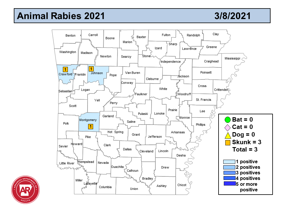
AR Rabies Map – Arkansas Public Health Veterinarian – Dr. Laura Rothfeldt
|
|
Report all animal bites or contact with wild animals to the nearest, local Arkansas Department of Health Unit.
|
|
Veterinary Continuing Education Opportunities – License renewal period starts now.
|
|
Arkansas Department of Agriculture
|
|
|
|
|
|
|
|