EXTRA EXTRA, READ ALL ABOUT IT!
SCIENCE DISCOVERS NEW ORGAN!
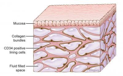
Some of you might have the seen the whirlwind of coverage across all media outlets on Wednesday about what they described as the discovery of a new organ (illustrated above left) lying just underneath our skin. This organ was described as “d
emonstrating that it is part of the submucosa and a previously unappreciated fluid-filled interstitial space, draining to lymph nodes and supported by a complex network of thick collagen bundles” in the paper published in the most recent issue of the journal
Scientific Reports
.
Wait, wait, hold on… did they just say a fluid filled region marked by collagen bundles? So you’re saying that science has just discovered FASCIA! Yes, that’s right, the amazing discovery by researchers out of Mount Sinai and The University of Pennsylvania is what John Barnes has been discussing for over 40 years. The same tissue that Dr. Jean-Claude Guimberteau was filming since at least 2001 and published in his films:
Strolling Under The Skin, Muscle Attitudes, The Skin Excursion
and
Interior Architectures
as well as his textbook,
Architecture of Human Living Fascia: The Extracellular Matrix and Cells Revealed through Endoscopy
(illustrated above right). Finally, this is the same tissue that has been researched by an ever-growing multitude of researchers culminating in the first International Fascia Research Congress, which will be in it’s 5
th
incarnation in 2018. But all of you already knew that, since we talk about it EVERY SINGLE SESSION, right?
So, as a celebration of science catching up, we will again share our 3-part series, first shared in 2015, on Fascia; which reads all the better with this current discovery. We hope you enjoy reading about more of the amazing properties of this “unrecognized interstitium”!
Benias, Petros C., et al. “Structure and Distribution of an Unrecognized Interstitium in Human Tissues.”
Scientific Reports
, vol. 8, no. 1, 2018, doi:10.1038/s41598-018-23062-6.
Guimberteau, J. and Armstrong, C. (n.d.). Architecture of human living fascia. Edinburgh: Handspring Publishing Limited.
Fascia: "What We Do Know"
Part 1 of a 3-Part Series
It is an unfortunate, yet incontrovertible, fact that medical and allied health programs have taught, and continue to teach, an incomplete model of the human body. This incomplete model that reflects a fragmented body made up of separate systems (cardiovascular, neurological, pulmonary, musculoskeletal, etc.), while impressive in its ability to understand and treat these separate systems, is inconsistent with the experience that we all have of a life in totality. While this concept of integration sounds logical and resonates with our intuitive understanding of life; to be accepted as an update to our current understanding, must have its basis in scientific fact. A cursory review of the fascia (pronounced fa-sha), or connective tissue system of the body, allows us to see the connection point between our current model of fragmentation and our updated model of integration.
As our previous statement alluded, the fascial system of the human body is largely ignored in the medical and allied health programs; and, subsequently, in our current healthcare system. The idea that a complete system of the human body should be overlooked becomes even more alarming when we realize that there exists over sixty years of research that contradicts the current model. The articles in this series will introduce the anatomical and physiological aspects of the fascial system that will allow us to appreciate the importance of this system of the body in truly understanding health and disease; as well as the great disservice medicine has done by ignoring it.
While the fascial system is largely overlooked in our medical training, certain facts are understood by the majority of healthcare practitioners. 1) The fascial system is an uninterrupted tissue that weaves its way through the body, surrounding, interpenetrating and connecting every single structure; and, 2) Fascia is composed of three components; the fibrous protein collagen, the stretchy fibers known as elastin and the gelatinous material that holds this colloidal structure together; the ground substance, sometimes referred to as the extracellular matrix. Unfortunately, medical dissection labs are often punctuated with the instruction to “scrape away all that unimportant tissue to get to the important stuff!” This crude understanding of fascia leads to the practice of relegating its function to that of biologic packing material; inert and passive, the Styrofoam peanuts of the body.
Firstly, let’s review the idea that there exists a tissue that is uninterrupted, much like our skin is uninterrupted, and that it weaves its way through the body connecting every single structure and cell to every other structure and cell. We might begin to envision a structure that resembles a spider’s web; and we would be closer to the reality of the situation. This web of tissue creates the shape and structure of our body, to the point where noted author and physical therapist John Barnes has stated that if every structure were removed from the human body aside from the fascial system, our shape would remain exactly the same 2 . This global understanding of fascia still leaves it as inert and passive; but why does this matter? Bioengineer Dr. Stephen Levin has shown how our current “post and beam” concept of the skeletal system and the further involved “lever and pulley” model of the musculoskeletal system are insufficient in explaining the dynamic support and structure of the human being 9 . Going further, researchers Dr. Robert Schleip 10 and Dr. Thomas Findley 5 have shown, respectively, that the fascia is capable of contracting, similarly to smooth muscle, as well being the transmitter of mechanical forces through the human body. This research reveals a tissue that is anything but passive.
We will now introduce a concept known as mechanotransduction to illustrate how this active tissue is involved in more than just our structure and our movement. Mechanotransduction is the phenomenon wherein mechanical signals outside of the cell are translated into biochemical signals inside the cell, including the nucleus. It is an accepted fact that nothing touches the outside of each cell except the ground substance of the fascial system; hence it being referred to as the extracellular matrix. Pioneering work by Drs. Ingber and Alenghat 1, 7 reveals the ground substance of the fascial system to be one of the main elements responsible for the phenomenon of mechanotransduction; leading to an understanding that this global web of tissue is involved in cellular function as well, including the very act of DNA transcription; the blueprint of our entire existence 7 . Research teams from the University of California – Berkeley have shown repeatedly that this mechanotransduction is directly affected by the viscosity of the ground substance. This research pointed to the effects that this viscosity had upon the incidence, as well as remission, of certain cancers 4, 13. This viscosity of the ground substance leads to another physiological aspect of the fascial system, phase transition. Phase transition is the scientific phenomena of matter changing from one state to Page 1 of 2 another; the example often used of ice changing to water, then to steam, then back again. Due to the fascia being colloidal (a solution wherein the particles are dispersed and do not sink), this phenomenon of phase transition is known as thixotropy; a term used to describe a colloidal gel-like substance, like fascia, that will undergo phase transition when stressed for a long enough duration 3 .
Our investigation has led us to understanding the fascia as an active, dynamic and adaptive tissue that responds to its environment mechanically. But what of the current assumption that fascia is inert? Nobel recipient Dr. Szent-Gyorgi postulated in a presentation in 1941 that the collagen in connective tissue, due to its arrangement molecularly, would act as a semiconductor 12; the same type of electrically conductive material that powers our modern day cell phones and computers. This hypothesis was later proved in experiments led by Dr. Szent-Gyorgi 6 . Further researchers discovered that, due to the fascia’s molecular arrangement being similar to crystals, would also possess the capacity for piezoelectricity (peezo-electricity). A piezoelectric material is any that, when subjected to mechanical stresses, will produce an electric charge 11. This has led noted fascial researcher, Dr. Helene Langevin to hypothesize that the fascial system could act as a body-wide communications array 8 .
The current understanding of the fascial system is of a passive and inert tissue that serves the role of glorified packing material. Conversely, research spanning sixty years has revealed a tissue that is contractile, uninterrupted, dynamic, adaptive and electrically active. Further, this tissue is the connection between every single one of our approximately 37 trillion cells and is partially responsible for the functions of each of those cells, including DNA transcription. So the next time that one of the therapists at the Inner Circle Myofascial Release and Physical Therapy Centers talks to you about the importance of fascia, you will begin to hear it through new ears. The therapists at Inner Circle have spent countless hours refining their ability to work with the fascial system through their expertise with skilled manual therapy techniques and have become Bucks County’s leading specialists and authorities on the John F. Barnes Myofascial Release Approach.
Part Two of this series will delve deeper into the complete integration of the human body via the fascial system, through the landmark research of Dr. Jean-Claude Guimberteau.
Professionally yours,
David Noonan PTA, BA
Expert Myofascial Release Therapist
References
1. Alenghat, F. and Ingber, D. (2002). Mechanotransduction: All Signals Point to Cytoskeleton, Matrix, and Integrins. Science Signaling, 2002(119), p6
2. Barnes, J. (1990). Myofascial release, the search for excellence. Paoli, Pa. (10 S. Leopard Road, Suite One): Myofascial Release Seminars.
3. Barnes, H.A. (1997). Thixotropy – A Review. Journal of Non-Newtonian Fluid Mechanics, 70(1997)1-33
4. Chaudhuri, O., Koshy, S., Branco da Cunha, C., Shin, J., Verbeke, C., Allison, K. and Mooney, D. (2014). Extracellular matrix stiffness and composition jointly regulate the induction of malignant phenotypes in mammary epithelium. Nat Mater, 13(10), pp.970-978.
5. Findley, MD, PhD, T. (2011). Fascia Research from a Clinician/Scientist’s Perspective. International Journal of Therapeutic Massage and Bodywork, 4(4). =.
6. Gascoyne, P., Pethig, R. and Szent-Gyorgyi, A. (1981). Water structure-dependent charge transport in proteins. Proceedings of the National Academy of Sciences, 78(1), pp.261-265.
7. Ingber, D. (2006). Cellular mechanotransduction: putting all the pieces together again. The FASEB Journal, 20(7), pp.811-827.
8. Langevin, H. (2006). Connective tissue: A body-wide signaling network? Medical Hypotheses, 66(6), pp.1074-1077.
9. Levin, S. (1995). The Importance of Soft Tissues for Structural Support of the Body. Spine: State of the Art Reviews, 9(2).
10. Schleip, R., Klingler, W. and Lehmann-Horn, F. (2007). Fascia is able to contract in a smooth muscle-like manner and thereby influence musculoskeletal mechanics.
11. Shamos, M. and Lavine, L. (1967). Piezoelectricity as a Fundamental Property of Biological Tissues. Nature, 213(5073), pp.267-269.
12. Szent-Gyorgi, A. (1941). Towards a New Biochemistry.
13. Yang, S. (2015). To revert breast cancer cells, give them the squeeze. [online] Newscenter.berkeley.edu. Available at: http://newscenter.berkeley.edu/2012/12/17/malignant-breast-cells-grow-normally-when-compressed/ [Accessed 22 Apr. 2015].