Targeting TrkC: Biosensis TrkC Antibodies in Action

TrkC (tropomyosin receptor kinase C) is a neurotrophin-3 (NT-3) receptor and a key target in neuroresearch due to its critical roles in neural development, synaptic plasticity, and neuroprotection. It regulates neuronal differentiation, axon guidance, and survival through pathways such as MAPK/ERK and PI3K/AKT. TrkC is studied in the context of neurodevelopmental disorders (e.g., autism, schizophrenia), neurodegenerative diseases (e.g., Alzheimer’s, Parkinson’s), and injury repair, where it supports neuronal regeneration and functional recovery. Its expression patterns and signaling dynamics make it an important biomarker and therapeutic target for understanding and treating various neurological conditions.

Biosensis Anti-TrkC antibodies

Biosensis Antibodies targeting TrkC provide precise detection and modulation of TrkC signaling. These antibodies are invaluable in studying neurodevelopment, disease progression, and potential interventions across a wide range of neurological disorders. Our antibodies are optimized for IHC/ICC, IF, Western Blot, ELISA, and Flow Cytometry.


Mouse Monoclonal Antibody to TrkC - M-1837-100

This antibody targets the extracellular domain of human TrkC and is suitable for applications like Flow Cytometry (FC) and Immunocytochemistry (ICC). It is cross-reactive with rodent species and is available in an unconjugated form.


Rabbit Polyclonal Antibody to TrkC (Whole Serum)-R-151-100

This antibody is designed for Immunohistochemistry (IHC) on frozen tissue sections. It demonstrates specificity for human, mouse, and rat TrkC and is produced using the extracellular domain of glycosylated human TrkC protein. 


Rabbit Polyclonal Antibody to TrkC (Affinity purified) - R-1835-100

This antibody is developed for use in Western Blotting (WB) applications. This antibody exhibits specificity for TrkC in human, mouse, and rat samples and is generated using a synthetic peptide immunogen from the ECD of human TrkC. The antibody exhibits no cross-reactivity to TrkA or TrkB by Western Blot.

Mouse Monoclonal Antibody to TrkC - (M-1837-100)

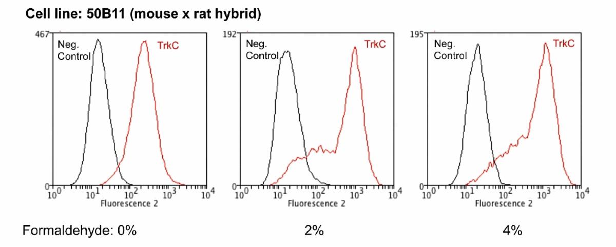
TrkC expression (red line) in 50B11 mouse x rat hybrid cell line analysed by Flow Cytometry using M-1837-100. The antibody is capable of detecting TrkC expression in unfixed as well as fixed conditions. The Flow cytometry data and results were generated using the Orflo Moxiflow™ instrument and protocols.

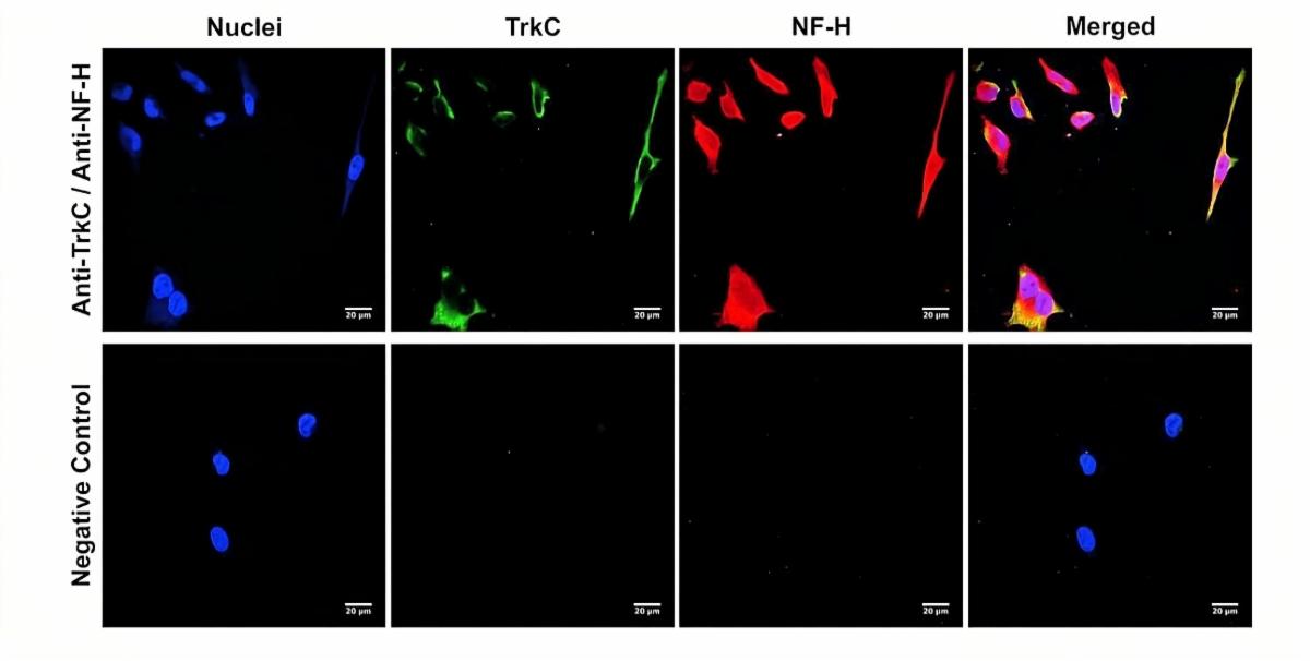
Membrane TrkC expression (green) in human HeLa cells, analyzed by Immunocytochemistry using M-1837-100 and colabelled with NF-H (red) by R-1389-50. Specific staining is observed for TrkC in the cell membrane, while the NF-H antibody reveals intracellular filaments.

Tyrosine Kinase Receptor C (TrkC), Rabbit Polyclonal Antibody (R-1835-100)

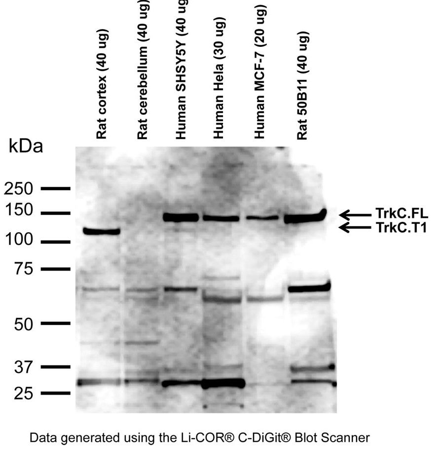
Western Blot analysis of TrkC expression in various human and rodent tissues and cell extracts (RIPA) using R-1835-100. Two prominent bands are detected at ~140 kDa and ~100 kDa, corresponding to TrkC full-length (FL) and truncated TrkC (T1) isoforms. Additionally, uncharacterized lower molecular-weight bands are observed as suspected degradation fragments.

Biosensis is a company that is passionate about Science

Email  LinkedIn  Web