 December
2018
December
2018
Contents
MSA Headquarters Address:
11130 Sunrise Valley Drive, Suite 350
Reston, Virginia 20191
|

M&M 2019
|
|
MEETING UPDATE
If you submitted a paper in 2018, your Catalyst login should still work. Or you can create a new account if you wish.
The deadline to submit is: February 15, 2019. See your November
Microscopy Today for a printed Call for Papers brochure and poster.
Don't forget to hang up your beautiful M&M 2019 poster in your office or lab!
|
 |
|
Association News
|
|
Cast Your Vote in the MSA Elections
As an MSA Member, the annual election of leadership is your opportunity to have an impact on the future of your Society. The MSA Nominations Committee has put together an impressive slate of candidates for the 2019 Election. The ballot can be accessed by logging into the Members Portal on the MSA website or by visiting
MSA Elections. Each candidate has submitted a brief bio, along with their credentials and their MSA Platform Statement. Please take a moment to review this information carefully. The voting process should take less than five minutes to complete.
Votes must be cast online by December 15.
Annual Undergraduate Research Scholarship Program
Applications are still being accepted for MSA's Annual Undergraduate Research Scholarship Program. The Scholarship Program is intended to foster the educational and research potential of full-time undergraduate students interested in pursuing microscopy as a career or major research tool. Applications for research involving a
ny area of microscopy are suitable for the program.
The deadline to apply is December 31
. Please visit the
MSA website
for further details.
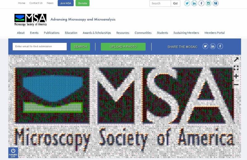
The Piece of the Whole MOSAIC
MSA is proud to introduce its newest collaborative project. Introducing the "Piece of the Whole" MOSAIC, a design to connect the society through visual media. The MOSAIC is a digital platform for you to share your microscopy and microanalysis work on the MSA homepage. It is through your eyes that we can focus the big picture of the society. Share your world within our world!
- Click the Upload Photo button at the top of the MOSAIC
- Search names, techniques and subjects to connect with other members of the society
- Cultivate the community and share the MOSAIC
|
|
|
Science News
|
|

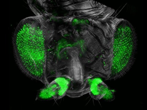
Advances in cellular microscopy: Transparent fruit flies
 |
|
|
An ultramicroscope picture of the head of Drosophila melanogaster. Credit: TU Wien |
A new kind of microscope has been developed: it creates 2D light sheets, penetrating biological tissues and causing special molecules to fluoresce. For the microscope to work, the tissue has to be made transparent, so the researchers developed new chemical procedures to 'clear' them. With this technique, extremely detailed pictures of intricate features of the nervous system of fruit flies are now possible. Read more here.
Making X-ray microscopy 10 times faster
Microscopes make the invisible visible. And compared to conventional light microscopes, transmission x-ray microscopes (TXM) can see into samples with much higher resolution, revealing extraordinary details. Researchers across a wide range of scientific fields use TXM to see the structural and chemical makeup of their samples-everything from biological cells to energy storage materials. Read more
here.
|
|
| Microscopy Today |
|
Microscopy Today
During the course of a year, many significant and interesting micrographs are acquired as still images or video clips. The editors of
Microscopy Today
wish to promote and honor such micrographs.
About a year ago, Editorial Board Member Robert Simmons and his wife Camille came to me with an idea. They suggested that
Microscopy Today
sponsor a micrograph competition where the scientific significance of an image weighs as much as the aesthetic qualities of the image. They volunteered to help implement such a contest. The MSA Council approved the project at M&M 2018 in Baltimore, and the
Microscopy Today
Micrograph Awards program is now set to begin, with the first awards to be presented at M&M 2019 in Portland, OR.
All types of microscopy are welcome. There are three micrograph categories:
Published micrograph category
for images appearing for the first time in a publication of the previous year. Thus, for the 2019 awards, only micrographs published in 2018 would be eligible.
Open micrograph category
for unpublished images acquired by any microscopist.
Video micrograph category
for movie clips up to 60 seconds in length, acquired directly with a microscope or generated as a video reconstruction from microscopy data.
A panel of judges will select a number of finalists in each category. Finalist micrographs will be displayed on social media, websites, and publications. From these finalists the judges will select a first prize winner in each category. The microscopy community at large will have an opportunity to vote for the "people's choice award," their choice for the best micrograph of the combined categories.
Submission of still images and videos for the
Microscopy Today
Micrograph Awards program will be through an MSA website available in January. The timeline begins with the submission deadline of February 21, 2019. Finalists will be notified in April. The
Microscopy Today
Micrograph Awards will be presented on Wednesday of the August M&M meeting. The top winners will receive cash prizes, and winning images will be featured on the cover of the September issue of this magazine.
I invite you to participate in this competition. Please submit your still images or videos. We look forward to receiving your work by the submission deadline of February 21, 2019.
Charles Lyman
Editor-in-Chief
Microscopy Today
|
| MSA Student Council News |
|
The MSA Student Council (StC) is organizing the 3rd annual Pre-meeting Congress for Students, Postdocs and Early-career Professionals in Microscopy & Microanalysis (PMCx60), to be held on the Saturday preceding M&M 2019 in Portland, OR.
The PMCx60 is a one-day conference organized by and for students and early-career professionals and offers a highly interactive forum for participants to share cutting edge research, network, and engage with peers ahead of the main meeting.
The StC is soliciting financial support from the MSA community and welcomes corporate sponsorship and donations from individuals/organizations to help build the future of microscopy and microanalysis.
Please consider sponsoring, donating to, and attending the 2019 PMCx60 in Portland, OR. To learn about the benefits of being a PMCx60 sponsor, email the StC at
[email protected]
, or donate to the PMCx60 via the MSA donation page (
microscopy.org/donation
, include "PMCx60" in the "Donation Made by:" field).
|
|
| Local Affiliated Societies |
|
Local Affiliated Societies News
by Patty Jansma, LAS Director
LAS Meetings
Check the individual LAS websites for more details.
Support your local affiliated society! Invite students, early career scientists, and technologists to your LAS meetings. Better yet,
bring a new member to your local meeting and get them involved!
LAS Programs
MSA provides LAS support with Tour Speakers, Grants-in-Aid, and Special Meeting grants. Details can be found at
http://www.microscopy.org
/communities/programs.cfm. Please contact me as soon as possible if your LAS is interested in applying, and you haven't already received funding this year.
As always, you may contact me at
[email protected] with comments, questions, or concerns.
Thanks!
Patty
|
|
| Focused Interest Groups |
|
Did you know that there are 11 FIGs that promote a specific discipline to microscopy? Click
here
to check them out and contact the leader to get more information.
You can join a FIG when renewing your MSA membership or while updating your member profile; students may join one FIG at no cost.
|
|
| In Memoriam |
|
 MSA announces with regret the passing of Sir Aaron Klug on November 20, 2018, at age 92. Klug was born in Lithuania, but grew up in South Africa. He received the Nobel Prize in chemistry in 1982 for the development of electron crystallography. After early work in metallurgy at Cambridge, in the 1950s and 1960s, he worked with Rosalind Franklin on the structure of Tobacco Mosaic Virus (TMV), and then worked on viruses with Kenneth Holmes and John Finch. He joined the MRC lab in Cambridge in 1962, and he was knighted in 1988. In 1970, his work with DeRosier and Crowther on 3-D reconstruction from projections resulted in a landmark paper in structural biology. MSA recognizes many Nobel prizes having been awarded to both light and electron microscopists.
|
|
MSA Job Placement Services
|
|
|
|
|
|
|
|