Looking for a specific section? Find it quick with these links below!
|
 |
|
Book your hotel room now! Special rates at the hotels below are available until July 1, 2020 or until the room blocks at the conference hotels are sold out.
Mark your calendars! Registration for M&M 2020 will open in March! Get a glimpse of what you can expect by viewing the Meeting Update where you'll find plenary sessions and program highlights, registration fees, and special M&M hotel rates.
The M&M 2020 Submission Site is still accepting abstracts!
Visit the
M&M 2020 website
and click the "Submit Your Paper button", or go directly to the submission site:
Submission deadline is Friday, February 21, 11:59 PM, U.S. Pacific Time.
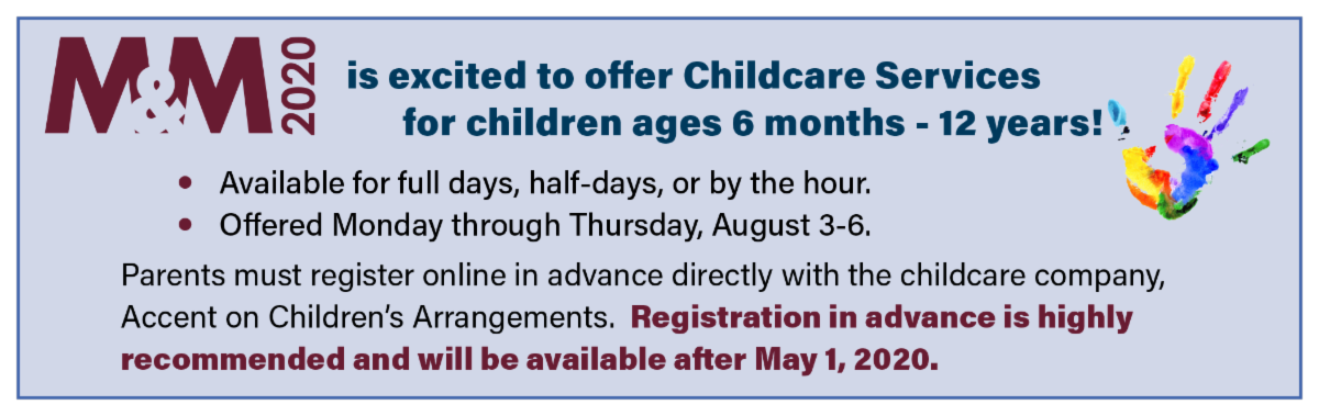
For the second year, M&M 2020 is excited to offer Childcare services for children ages 6 months - 12 years. Services will be available Monday through Thursday, August 3-6 for full days, half-days, or by the hour. Registration in advance is highly recommended and will be available after May 1, 2020.
|
|
|
Do you ever wonder how people get chosen to walk across the stage at M&M to receive an award? Here are some tips and tricks that you need to know!
|
|
|
Pictured above: Biological Sciences Poster Winner, Areen Badwal with Past-President Paul Kotula at M&M 2019. Photo credit: Dallager Photography
|
|
|
|
|
Pictured above: Analytical Sciences Poster Winner, Arthur Moya with Past-President Paul Kotula at M&M 2019. Photo credit: Dallager Photography
|
|
Tips and Tricks for a winning student poster award at M&M:
- Read and follow instructions for preparation of your poster
- Use the large poster boards wisely by creating BIG images (with mag bars, and figure legends)
- Create large drawings, figures and tables so that they are readable from several feet away from the poster
- Colors are good
- Be present at your poster so that you have a chance to talk with the judges
- Have a 3 minute elevator speech prepared to share with the judges
- Show data and conclusions
- Tell your research story, who, what, why
Judging criteria for the Ultramicrotomy Award by Diatome:
- Posters hanging in the M&M meeting exhibit hall all week by any members or attendees as judges will be on the lookout all week for great images
- In Materials and Methods: state that you used a diamond knife to prepare your sample by Ultramicrotomy (diamond knives inside an SEM are not eligible)
- Large, high resolution images
- Excellent composition, contrast and focus for microscope images of samples cut with diamond knife on an ultramicrotome
- Sample preparation will affect the images and results
_____________________________________________________________________
Submit an Article to Microscopy Today
The Editors of
Microscopy Today
(MTO) encourage and greatly appreciate submission of articles from microscope users as well as microscope manufacturers and suppliers. Of particular interest are summaries of in-depth articles published in peer reviewed journals and articles that describe new equipment and applications.
Microscopy Today
is open access and there are no charges for publishing in MTO. All articles are available free to our subscription list of over 18,000 microscopists and through our collaboration with Cambridge University Press over 8,000 libraries worldwide. For further information email the Editor-in-Chief at [email protected]
or visit
_____________________________________________________________________
Do you have a colleague that takes great micrograph images or video?
Share the information below!
|
|

|
|
 The MSA Facebook page regularly posts science news for you
Marriage of Microscopy Techniques Reveals Cells' 3D Ultrastructure
Scientists at the Howard Hughes Medical Institute (HHMI) have combined superresolution fluorescence microscopy and electron microscopy to shed new light on the structures and organizations within cells. Superresolution fluorescence microscopy on its own is able to show cellular structures with a high degree of clarity and detail, though the technique does have its limits. Only a few of the more than 10,000 proteins in a cell can be revealed, which does not provide researchers with enough information to study the interconnections and dependencies of cellular structures.
Read more here
.
Live-cell Imaging: Polarization adds rich context to SIM images
Polarization, a fundamental physical characteristic of light, has proven useful for various applications. In biology, polarization imaging reveals the orientation of targeted proteins by measuring the dipole orientation of fluorophores. Although superresolution polarization imaging has been used increasingly to study protein orientation, most such techniques are limited to labs with specialized expertise in these methods. Now, however, standard commercial structured illumination microscopy (SIM) systems can achieve superresolution polarization imaging, thanks to the work of researchers led by Peng Xi at Peking University and Qionghai Dai at Tsinghua University (both in Beijing, China). Read more here.
|
|
|
|
Share your Images with the Community!
Want the world to see your awesome images? Send them to us to be featured on the MSA StC #MicroscopyMonday posts. Click the link below to submit your image and follow us on Instagram @msastudentouncil to keep up with the latest news.
2020 Webinar Series
Next Webinar
: First week of March
Topic
: Core Facilities: what is available and how to apply
We are excited to announce that each student who attends the remaining webinars in this year's series will be entered to win a $25 Amazon gift card! Make sure you sign up for this upcoming webinar to receive more information on this opportunity.
Did you miss the last webinar about Communicating Science? No worries! Watch the full webinar (
https://youtu.be/UA-45wKMIj8
), subscribe to our YouTube channel, and join our future webinars.
Future Webinar Topics
Applications Scientist: Career options in Industry
Basic Microscopy Techniques
Preparing for M&M
Electron Microscopy Summer Schools 2020
Main courses at the Lehigh Microscopy School include a variety of SEM and TEM techniques. Specialized Courses cover everything from FIB to quantitative X-ray Microanalysis. See more details about the available courses
here
.
--------
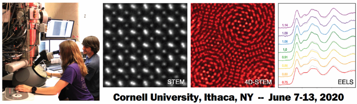
From June 7-13, 2020 NSF innovation platform PARADIM will host the 2nd Summer School on Electron Microscopy. The School will cover 2D, 3D and 4D imaging in the scanning transmission electron microscope (STEM), and will explore:
- fundamental and practical aspects of aberration correction
- electron energy loss spectroscopy (EELS)
- spectroscopic mapping
- cryogenic STEM
- imaging using pixelated detectors
The Summer School is divided into morning lectures and afternoon hands-on labs on state-of-the-art scanning transmission electron microscopes as well as on simulation and analysis software.
Application Deadline
: February 15, 2020
|
|
Local Affiliated Societies
|
Local Affiliated Societies News
by Patty Jansma, LAS Director
LAS Meetings
Check the individual LAS websites for more details.
Support your local affiliated society! Invite students, early career scientists & technologists to your LAS meetings. Better yet,
bring a new member to your local meeting and get them involved!
LAS Programs
As always, you may contact me at
[email protected]
with comments, questions or concerns.
|
|
|
|
|
Renu Sharma, Chair
Join a FIG
! FIGs are groups of scientists that practice or have interests in specific disciplines (currently 11) to which microscopy and microanalysis is applied. As an MSA member, you can join one or more (FIG Communities). FIGs not only boost scientific understanding through knowledge sharing, but also provide opportunity to network with scientists who share common interests. FIGs may organize lunches, symposia or pre-meeting congresses at M&M. A complete list of FIGs is on MSA website or by clicking
here
. You may contact the FIG leader directly or attend a business meeting at M&M to learn more. Visit the
FIG Store
to sign up. Are you already a member of a FIG? Consider volunteering and make an impact! It's members are what makes FIGs successful. Talk to your FIG leader. Interested in starting a FIG? Start by reviewing the updated version of the FIG Guidelines and then contact me. FIGs are for students too! If you are a student, your fees are waived for the first FIG you join.
Are you interested in highlighting your FIG in an MSA Update?
Contact me
for more information.
The Diagnostic & Biomedical FIG is organizing a Premeeting Congress (PMCX62), "
Contemporary Electron Microscopy Advances in Biomedical Research
", at M&M 2020 in Milwaukee.
Meetings sessions include:
- Automation and streamlining of workflow for diverse EM specimens
- Immuno electron microscopy and correlative LM-EM workflow, advances and challenges
- Challenges and Best practice in the preparation of plant, insect, aquatic/marine, and pharmaceutical specimens for EM
- Round table discussion: instrument-assisted and automated Bio EM sample processing, challenges and applications
Please click
here
for more details, including information about submitting an abstract.
|
|