Looking for a specific section? Find it quick with these links below!
|
 |
|
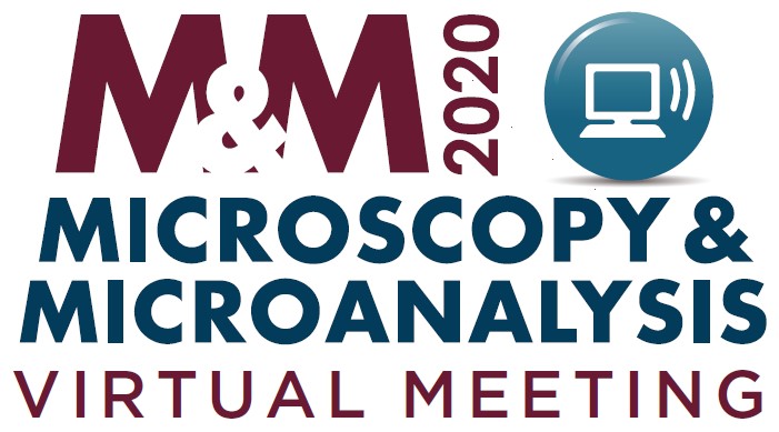
M&M 2020 Virtual is only weeks away!
M&M 2020 Virtual is quickly approaching and we're gearing up for an exciting new way to experience all the conference has to offer.
Later this month, we will be releasing a few quick tutorials to help you get acquainted with our virtual platform. Stay tuned for more information. In the meantime, here are some answers to frequently asked questions and the full scientific program
with all the educational content you can experience when you register for M&M 2020 Virtual today.
- FAQS: Click HERE for a list of FAQs about the virtual meeting and platform.
- Schedule at a Glance: View the week at a glance HERE.
- Scientific Program: To view the Full Scientific Program Listing, including all Platform and Poster presentations, click HERE.
- Registration: Click HERE to Register! For more details, visit the Registration Fees and Information page.
Join us August 3-7 for live and pre-recorded talks, live text and video chats with presenters, exhibitor demos and tutorials, product information, and vendor networking, two Pre-Meeting Congresses, 24/7 access to the meeting content, and unlimited opportunities to have private video chats with other attendees.
(Separate registration required for each individual participant.)
|
Non-Member
Rate
|
MSA/MAS
Member Rate
|
Student/Post-doc/ Emeritus Rate
|
| $150.00 |
$75.00 |
$20.00 |
|
|
|
Nominations for MSA Fellows, Class of 2021 is Now Open
The Microscopy Society of America is accepting nominations for MSA Fellow, Class of 2021.
This major Society award honors MSA members of at least 10 years for "
significant contributions to the advancement of the field of microscopy and microanalysis"
through both their scientific accomplishments AND service to MSA. In very special circumstances the 10 year membership requirement may be waived. Self-nominations will not be accepted.
The Society encourages all of its members to participate in honoring eligible colleagues in the field of microscopy by nominating them for MSA Fellow. The deadline is September 30, 2020. For instructions on the nomination process and submission of a nomination, please click
here
.
Submit an Article to Microscopy Today
The Editors of
Microscopy Today (MTO) encourage and greatly appreciate submission of articles from microscope users as well as microscope manufacturers and suppliers. Of particular interest are summaries of in-depth articles published in peer reviewed journals and articles that describe new equipment and applications.
Microscopy Today is open access and there are no charges for publishing in MTO. All articles are available free to our subscription list of over 18,000 microscopists and through our collaboration with Cambridge University Press over 8,000 libraries worldwide. For further information email the Editor-in-Chief at
[email protected] or visit
Did you have time to complete the crossword puzzle on pages 68-69 of the May issue of
Microscopy Today
? Click the puzzle below for an interactive version!
Theme: Light Microscopy b
y Erica Clark and Aubrey Penn
Answers to the crossword puzzle can be found on Page 81.
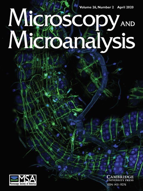
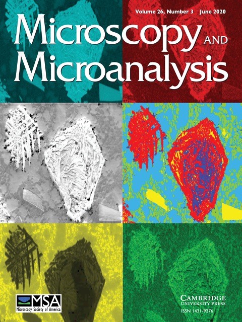
Microscopy and Microanalysis Journal
The April 2020 and June 2020 issues of the
Microscopy and Microanalysis Journal is being sent out as a printed copy to U.S. subscribers. Be on the lookout for your copies in the mail!
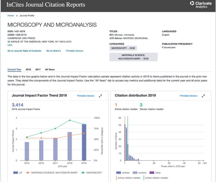
In case you missed it, the
Microscopy and Microanalysis Journal's New Impact Factor is 3.414, up from 2.673 last year. Take a look at the stats below provided by Editor, John Mansfield.
|
|
|
You may have recently seen the Center for Open Bioimage Analysis (COBA
) name and logo around various parts of the image.sc forum
; we are a new center from Kevin Eliceiri (UW-Madison) and Anne Carpenter's (Broad Institute) labs whose mission is to serve the biology community's growing need for sophisticated software for light microscopy image analysis. We will do this in two main ways:
- In collaboration with biological partner labs, we will create new tools and improve existing tools to make new analyses possible and existing analyses easier.
- We will do outreach to the biology community to help spread knowledge on how to best apply bioimage analysis tools and approaches.
In order to do both of these well, we need a representative picture of the kinds of images our community is analyzing, what they think is working well, what they think is NOT working well, and what sorts of outreach and educational materials they prefer (in person, written, videos, etc).
To that end, we've written a brief 10-15 minute survey
(open until July 31st) to ask you, the community, what you work on and how you'd like to communicate and collaborate with us! We would especially ask you share this with your local microscopy and image analysis communities - the more representative of a sample we get of users, the better we'll serve the whole field.
If you want to learn more, please reach out to the COBA Team via
our website.
|
|
Upcoming Microscopy Today Webinar
|
|
Utilizing Dynamic CT Imaging for True In Situ Experimentation
Thursday, August 13, 2020 |
1:00 p.m. ET
The technique of time-resolved 3D imaging with X-rays has rapidly emerged as an essential part of a researcher's toolkit for understanding materials evolution. The expansion into "4D" has facilitated in situ investigations ranging from mechanical deformation of solids to fluid flow in porous materials to the evolution of structure in changing environmental conditions. Imaging of dynamic processes specifically processes happening over a short time-period, has been one of the key applications at synchrotron micro-CT beamlines, with temporal resolutions well below one second. However, access to these facilities is often limited, with respect to both time at the beamline and application specificity.
In the laboratory, image quality and spatial resolution have been significantly improved over the last decade, providing researchers routine and regular access to very high quality computed tomography data. However, the advances in temporal resolution have been less accelerated, especially for imaging dynamic processes. Lab-based CT has been limited to pseudo in situ experimentation where data are collected on interrupted processes allowing for the traditionally longer and non-continuous scanning protocols. Additionally, analyzing and visualizing complicated data from 3D studies has been fairly burdensome. Over the last several years, development at TESCAN has focused on addressing these problems and has now made it possible to acquire, visualize, and analyze micro-CT data on uninterrupted and dynamic processes. Through a number of hardware and software advances, temporal resolution has been pushed below 10 seconds per scan, opening up new frontiers for lab-based 4D CT work.
In this talk we explore the challenges and innovations that have led to dynamic CT, highlighting several applications across materials sciences, life sciences, and geosciences. Click here to register.
|
|

|
 The MSA Facebook page regularly posts science news for you
Faster processing makes cutting-edge fluorescence microscopy more accessible
Scientists have developed new image processing techniques for microscopes that can reduce post-processing time up to several thousand-fold. The researchers are from the National Institutes of Health with collaborators at the University of Chicago and Zhejiang University, China.
Read more
.
Electron cryo-microscopy: Using inexpensive technology to produce high-resolution images
Biochemists at Martin Luther University Halle-Wittenberg (MLU) have used a standard electron cryo-microscope to achieve surprisingly good images that are on par with those taken by far more sophisticated equipment. They have succeeded in determining the structure of ferritin almost at the atomic level. Their results were published in the journal PLOS ONE.
Read more
.
|
|
|
|
|
StC Executive Council Elections
MSA Student Council Announces Slated Executive Council Positions
MSA Student Council (StC) is pleased to announce Slate for the StC Executive Council (EC) Officer positions!
As per the
MSA Student Council Policies and Procedures, each MSA Student Member as of May 1st will receive an electronic ballot for the EC elections. Student Members will have from July 10th to July 30th to cast their vote for EC.
A description of the roles for the three executive positions (President-Elect, Secretary, and Treasurer) can be found on the MSA Student Council website,
Responsibilities page. The President-Elect serves a three-year term, one each as President-Elect, President, and Past-President while the Secretary and Treasurer positions serve a one-year term.
President-Elect: Aubrey Penn, North Carolina State University
 I am a third-year graduate student in Materials Science and Engineering at North Carolina State University working with Profs. James LeBeau and Divine Kumah. My research is focused on explaining interfacial phenomena in complex oxide thin films using scanning transmission electron microscopy imaging and spectroscopy. Before coming to NCSU, I received my master's degree in chemistry from Western Kentucky University, where I first became acquainted with electron microscopy. Since then I have grown in knowledge of the technique and love of the microscopy field, in part, through my association with MSA and MSA Student Council. I have been a member for three years, and served as Secretary this year. If elected, I will actively pursue diversity and inclusivity to strengthen MSA, increase outreach efforts worldwide through Regional Liaison involvement, and create an outlet for post-doctoral researchers to be more integrated into the Society. Additionally, I will work to maintain the welcoming community I have come to know and appreciate.
Secretary: Louisa Mezache, Ohio State University
 I'm a PhD student at The Ohio State University in Dr. Sai Veeraraghavan's lab, where I study the functional effects of structural remodeling of the cardiac intercalated disc. I was introduced to microscopy while working for a small start-up, but have gained a greater appreciation for it while working with Dr. Veeraraghavan. Prior to returning to school, I worked as a clinical research coordinator in neuromuscular diseases. There I helped patients with spinal muscular atrophy (SMA) access a novel millions-of-dollars drug treatment. I attended my first M&M in 2019 and joined the MSA Student Council as the Biological Sciences Co-chair, and I'm excited by the opportunity to continue participating. If selected, my intention will be to help broaden participation from untapped groups in MSA, both social and scientific, by increasing year-round engagements through social media. I'm a PhD student at The Ohio State University in Dr. Sai Veeraraghavan's lab, where I study the functional effects of structural remodeling of the cardiac intercalated disc. I was introduced to microscopy while working for a small start-up, but have gained a greater appreciation for it while working with Dr. Veeraraghavan. Prior to returning to school, I worked as a clinical research coordinator in neuromuscular diseases. There I helped patients with spinal muscular atrophy (SMA) access a novel millions-of-dollars drug treatment. I attended my first M&M in 2019 and joined the MSA Student Council as the Biological Sciences Co-chair, and I'm excited by the opportunity to continue participating. If selected, my intention will be to help broaden participation from untapped groups in MSA, both social and scientific, by increasing year-round engagements through social media.
Treasurer: Jackson Spurling, University of Tennessee - Knoxville
 The opportunities to serve as Region VIII Liaison during 2017-18, as Secretary during 2018-19, and as Treasurer during 2019-20 have allowed me to become involved with the MSA StC leadership, particularly in the area of member outreach. In these positions, I have assisted the MSA Student Council chiefly through its goal to provide networking platforms to student members. This has been accomplished through having dialogue with regional members as well as through activities like webinars and social media outreach. I would like to continue to contribute to the Student Council by supporting these and other methods of outreach to student members through the position of Treasurer. I have been honored to serve with StC these last three years, and I hope to be given the opportunity to continue to make MSA a welcoming organization for new and current members by supporting current and future outreach initiatives. The opportunities to serve as Region VIII Liaison during 2017-18, as Secretary during 2018-19, and as Treasurer during 2019-20 have allowed me to become involved with the MSA StC leadership, particularly in the area of member outreach. In these positions, I have assisted the MSA Student Council chiefly through its goal to provide networking platforms to student members. This has been accomplished through having dialogue with regional members as well as through activities like webinars and social media outreach. I would like to continue to contribute to the Student Council by supporting these and other methods of outreach to student members through the position of Treasurer. I have been honored to serve with StC these last three years, and I hope to be given the opportunity to continue to make MSA a welcoming organization for new and current members by supporting current and future outreach initiatives.
StC Appointed Position Applications Are Open!
The Call for Applications for the Appointed Positions (Program Chair, Communications Chair, Regional Liaison Chair, PMCx60 Co-chairs, and Regional Liaisons) are open. The application and role descriptions can be accessed on the MSA Student Council website,
Responsibilities
page. Application materials must be submitted by August 15th to [email protected].

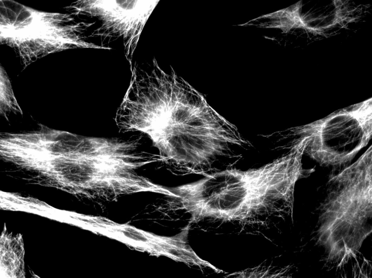
Sarah Kearns
 July's Student Spotlight is fifth-year graduate student and scientific writer, Sarah Kearns. Sarah works with Drs. Michael Cianfrocco and Kristen Verhey in the Program in Chemical Biology at the University of Michigan. Her project combines cell biology and structural biology to study issues in mitosis that can lead to cancer. While primarily a biochemist, she has worked with single particle cryo-TEM and fluorescence microscopy to characterize complex cellular structures involved in cell division. An example image from her research, shown below, is a fluoresce image of the microtubule network in cells. July's Student Spotlight is fifth-year graduate student and scientific writer, Sarah Kearns. Sarah works with Drs. Michael Cianfrocco and Kristen Verhey in the Program in Chemical Biology at the University of Michigan. Her project combines cell biology and structural biology to study issues in mitosis that can lead to cancer. While primarily a biochemist, she has worked with single particle cryo-TEM and fluorescence microscopy to characterize complex cellular structures involved in cell division. An example image from her research, shown below, is a fluoresce image of the microtubule network in cells.
Nearing the end of her graduate career, Sarah has a wealth of experiential knowledge for incoming graduate students. Her practical advice is first to keep a very detailed notebook. Once the thesis-writing stage arrives, remembering small details from an experiment 3-4 years ago is nearly impossible. She also encourages students to look for a group where they not only like the science, but also get along well with the lab.
 Sarah is quite involved with science writing as the Editor-in-Chief for Michigan Science Writers, an organization for training students and postdocs to write and edit for a general public about science. This group has produced EquilibriUM, a magazine to make science, technology, engineering, art, and medicine more engaging and accessible. Sarah also maintains a blog called
Annotated Science
. After graduation, Sarah will join the Knowledge Futures Group, working to curate and preserve knowledge for the good of the people. Sarah has additionally used her scientific communication savvy this summer helping MSA StC with our Social Media Subcommittee. Sarah is quite involved with science writing as the Editor-in-Chief for Michigan Science Writers, an organization for training students and postdocs to write and edit for a general public about science. This group has produced EquilibriUM, a magazine to make science, technology, engineering, art, and medicine more engaging and accessible. Sarah also maintains a blog called
Annotated Science
. After graduation, Sarah will join the Knowledge Futures Group, working to curate and preserve knowledge for the good of the people. Sarah has additionally used her scientific communication savvy this summer helping MSA StC with our Social Media Subcommittee.
Sarah also enjoys baking and photography in her free time, and has more recently taken up gardening and learning piano.
|
|
An example image from her research, a
fluoresce image of the microtubule network in cells.
|
2020 Webinar Series
Each student who attends the remaining webinars in this year's series will be entered to win a $25 Amazon gift card! Make sure you leave your email address with us during the upcoming webinars to receive more information on this opportunity.
Mark your calendars! MSA StC is excited to announce that our 2nd annual "Preparing for M&M" webinar will be hosted this
Thursday, July 16, at 4 pm Eastern Time
. If you're interested in this event, please remember to
register
and be sure to join us for the live webinar via Zoom (you will receive the link prior to the webinar). Our panelists will answer all your questions about how to make the most of your M&M experience this year! If you have any burning questions, please let us know by filling out our
question survey form
and we'll be sure to pose your questions to our panelists. If you can't attend the live event, be on the lookout for the recording to be posted to our YouTube channel. We look forward to seeing you on Thursday!
Did you miss the last webinar about Applications Scientists? No worries! Watch the full webinar (
https://youtu.be/rqslwWn4LgM
), subscribe to our YouTube channel, and join our future webinars.
Share your images with the community!
Want the world to see your awesome images? You could be featured on the MSA #MicroscopyMonday posts. Submit to the MOSAIC: click "Upload a Photo" at
https://www.microscopy.org/
.
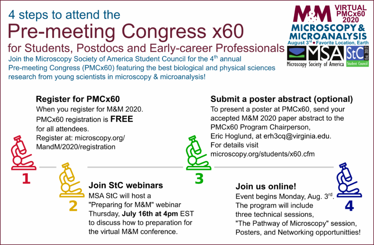
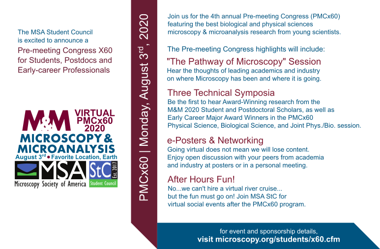
The MSA Student Council (StC) is organizing the 4th annual Pre-meeting Congress for Students, Postdocs, and Early-career Professionals in Microscopy & Microanalysis (PMCx60), to be held virtually on the Monday preceding M&M 2020.
The PMCx60 is a one-day conference organized by and for students and early-career professionals and offers a highly interactive forum for participants to share cutting edge research, network, and engage with peers ahead of the main meeting.
The StC is soliciting financial support from the MSA community and welcomes corporate sponsorship and donations from individuals/organizations to help build the future of microscopy and microanalysis.
Please consider sponsoring, donating to, and attending the virtual 2020 PMCx60.
To learn about the benefits of being a PMCx60 sponsor, email the StC at
[email protected], or donate to the PMCx60 via the MSA donation page (
microscopy.org/donation, include "PMCx60" in the "Donation Made by:" field).
For event details visit
https://www.microscopy.org/students/x60.cfm. Please email the PMCx60 Program Chair, Eric Hoglund, at
[email protected] if you are interested in sponsorship or have any questions about the PMC.
|
|
Local Affiliated Societies
|
Local Affiliated Societies News
by Patty Jansma, LAS Director
LAS Meetings
Check the individual LAS websites for more details.
Support your local affiliated society! Invite students, early career scientists and technologists to your LAS meetings. Better yet,
bring a new member to your local meeting and get them involved!
LAS Programs
As always, you may contact me at
[email protected]
with comments, questions or concerns.
|
|
|
|
|
|
Renu Sharma, Chair
Join a FIG
! FIGs are groups of scientists that practice or have interests in specific disciplines (currently 11) to which microscopy and microanalysis is applied. As an MSA member, you can join one or more (FIG Communities). FIGs not only boost scientific understanding through knowledge sharing, but also provide opportunity to network with scientists who share common interests. FIGs may organize lunches, symposia or pre-meeting congresses at M&M. A complete list of FIGs is on MSA website or by clicking
here
. You may contact the FIG leader directly or attend a business meeting at M&M to learn more. Visit the
FIG Store
to sign up. Are you already a member of a FIG? Consider volunteering and make an impact! It's members are what makes FIGs successful. Talk to your FIG leader. Interested in starting a FIG? Start by reviewing the updated version of the FIG Guidelines and then contact me. FIGs are for students too! If you are a student, your fees are waived for the first FIG you join.
Are you interested in highlighting your FIG in an MSA Update?
Contact me
for more information.
|
|