Looking for a specific section? Find it quick with these links below!
|
Time is winding down to save at the early bird rate!
Save on M&M 2021 Virtual when you register by Tuesday, June 15! We are excited to present a dynamic lineup of plenary speakers including, 2020 Kavli Awardee, Ondrej Krivanek, PhD, and COVID-19 vaccine developers, Kizzmekia S. Corbett, PhD and Jason McLellan, PhD. Join us for a robust scientific programming schedule along with live-chat Q&A sessions, opportunities to network, and so much more. Follow this link to reserve your virtual seat today:
https://www.microscopy.org/MandM/2021/registration/index.cfm.
|
M&M 2021 Post-Deadline Virtual Posters
DEADLINE APPROACHING!
Post-deadline posters may be submitted until the deadline of
Wednesday, June 30, 2021.
More information on submission requirements can be found in the link here.
|
|
 |
|
Create an Impact by Volunteering on an MSA Committee!
MSA Committees ensure the strength of our organization by promoting clear and concise goals for our volunteers. As a volunteer, the impact you create is the collaborative support of other committees to maintain and develop MSA as a whole. Take your membership to the next level by getting involved on one of our committees!

|
The MOSAIC is a digital platform for you to share your microscopy and microanalysis work on the MSA homepage. It is through your eyes that we can focus the big picture of the Society. Share your world within our world!
- Click the Upload Photo button at the top of the MOSAIC
- Search names, techniques and subjects to connect with other members of the society
- Cultivate the community and share the MOSAIC
|
Submit an Article to Microscopy Today
The Editors of Microscopy Today (MTO) encourage and greatly appreciate submission of articles from microscope users as well as microscope manufacturers and suppliers. Of particular interest are summaries of in-depth articles published in peer reviewed journals and articles that describe new equipment and applications. Microscopy Today is open access and there are no charges for publishing in MTO. All articles are available free to our subscription list of over 18,000 microscopists and through our collaboration with Cambridge University Press over 8,000 libraries worldwide. For further information email the Editor-in-Chief at Bob.Price@uscmed.sc.edu or visit
Submitted by John Mansfield, Editor, Microscopy and Microanalysis
Interested in publishing Open Access in Microscopy and Microanalysis?
Are you affiliated with an institution that is covered by one of CUP's Read and Publish agreements? If so, you may be eligible to publish your manuscript as Open Access at no cost to you.
There are agreements in place in over 42 countries around the world. The agreements have been established by many universities and consortia of universities around the world together with a number of national laboratories and research institutes.
For a full list of agreements, please view the R&P Agreements Page. You can also use CUP's checker tool, which will allow you to input your country and institution to pull up R&P eligibility status and terms.
If you wish to publish Open Access and are covered by one of these agreements, you will need to complete one of these forms and send it to our production team leader (Brian Mazeski - bmazeski@cambridge.org).
Publishing Open Access will improve people's ability to access your publication and, I believe, increase citations and also have a positive affect on our impact factor overall.
|
|

|
 The MSA Facebook page regularly posts science news for you The MSA Facebook page regularly posts science news for you
How Eikon Uses Fluorescence Microscopy for Drug Discovery
Fluorescence Microscopy is one of the most recent whereabouts in microscopy. In Fluorescence Microscopy, cells or tissues become stained with fluorophore, allowing it to emit colored light after being exposed to a certain wavelength. This can lead to better visualization of images of cellular objects. Read more.
Fluorescence Microscopy Peers Deep in the Brain
Optics & Photonics recently published a research journal about researchers developing a technique called, Diffuse Optical Localization Imaging (DOLI) to allow them to see blood flow without removal of the skull. This article was organized and edited by Mara Johnson-Groh. Read More.
|
|
|
|
|

Microscopy Career Miniseries:
Want to learn about career development in academia and national labs? Register to attend on Zoom or watch live on YouTube. Registrants can ask questions and network with professionals in the field.
|
Academia
Thursday, June 17 @ 1:00pm ET featuring
Prof. Beth Dickey, Carnegie Mellon University
Prof. Jinwoo Hwang, Ohio State University
Prof. Jim Howe, University of Virginia
|
National Labs
Tuesday, June 22 @ 12:00 PM ET
featuring
Dr. Steven Spurgeon, Pacific Northwestern National Laboratory
Dr. Juan Carlos Idrobo, Oak Ridge National Laboratory
Dr. Rhonda Stroud, US Naval Research Laboratory
|
5th Annual Pre-Meeting Congress for Students, Postdocs, and Early-Career Professionals (PMCx60)
On behalf of the PMCx60 organizing team and MSA Student Council, we are excited to announce this year's Pre-Meeting Congress, which will be held on July 31st. Don't forget to register for PMC while registering for the M&M 2021.
Ellen Zhong - Biological Sciences
 June's Biological Sciences Student Spotlight is a fourth year PhD student at Massachusetts Institute of Technology (MIT) in the Computation Systems Biology Program. She is co-advised by Drs. Bonnie Berger and Joseph Davis in this interdisciplinary program. Ellen's PhD research involves developing algorithms for cryogenic electron microscopy (cryo-EM) reconstruction, specifically using neural network machine-learning based algorithms to reconstruct dynamic protein structures. June's Biological Sciences Student Spotlight is a fourth year PhD student at Massachusetts Institute of Technology (MIT) in the Computation Systems Biology Program. She is co-advised by Drs. Bonnie Berger and Joseph Davis in this interdisciplinary program. Ellen's PhD research involves developing algorithms for cryogenic electron microscopy (cryo-EM) reconstruction, specifically using neural network machine-learning based algorithms to reconstruct dynamic protein structures.
 Ellen's first exposure to research and computation was as an undergraduate chemical engineering student at the University of Virginia (UVA), where she explored protein-folding processes via computational simulations. Her undergraduate research experience lead her to an internship in New York City with D. E. Shaw Research (DESRES), a company focused solely on developing super computers for running molecular dynamic simulations. Ellen continued working for the company for three years after graduating, an experience she found to be incredibly rewarding. As a young student, Ellen had always set her sights on graduate school, but the general knowledge she gained at DESRES better prepared her for her graduate studies. She had the time to figure out which field of research she wanted to pursue, and learned a healthy work-life balance that is certainly underrated in most graduate students' lives! Ellen's first exposure to research and computation was as an undergraduate chemical engineering student at the University of Virginia (UVA), where she explored protein-folding processes via computational simulations. Her undergraduate research experience lead her to an internship in New York City with D. E. Shaw Research (DESRES), a company focused solely on developing super computers for running molecular dynamic simulations. Ellen continued working for the company for three years after graduating, an experience she found to be incredibly rewarding. As a young student, Ellen had always set her sights on graduate school, but the general knowledge she gained at DESRES better prepared her for her graduate studies. She had the time to figure out which field of research she wanted to pursue, and learned a healthy work-life balance that is certainly underrated in most graduate students' lives!
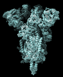 On her journey to identifying the field of study she would want to focus her PhD on, Ellen came across a paper by Roy Lederman, a mathematician at Yale University, who described the continuous heterogeneity problem in cryo-EM. That is, how do you reconstruct heterogeneous protein complexes and model continuous motion? This was the monster problem Ellen decided to take on for her dissertation! She went on to develop the software to model heterogeneity, and provide the software package for others to use. Though Ellen was very modest when sharing this accomplishment, she was excited to contribute something that the scientific community needed and could readily use. On her journey to identifying the field of study she would want to focus her PhD on, Ellen came across a paper by Roy Lederman, a mathematician at Yale University, who described the continuous heterogeneity problem in cryo-EM. That is, how do you reconstruct heterogeneous protein complexes and model continuous motion? This was the monster problem Ellen decided to take on for her dissertation! She went on to develop the software to model heterogeneity, and provide the software package for others to use. Though Ellen was very modest when sharing this accomplishment, she was excited to contribute something that the scientific community needed and could readily use.
Outside of her research, Ellen loves climbing, a hobby she found during her time in NYC. She also enjoys cooking and hosting dinner parties where she samples her new recipes.
Ellen has had such an impressive graduate career so far, and she is only getting started!
Gianluca Roscioli - Physical Sciences
 Congratulations to this month's student spotlight, Gianluca Roscioli. Gianluca is a PhD candidate in the Tasan group at MIT. He works on building in-situ setups in a scanning electron microscope (SEM) to understand the degradation mechanisms of metals. Congratulations to this month's student spotlight, Gianluca Roscioli. Gianluca is a PhD candidate in the Tasan group at MIT. He works on building in-situ setups in a scanning electron microscope (SEM) to understand the degradation mechanisms of metals.
Originally from Rome, Italy, he received his BSc and Msc at Politecnico di Milano in Materials Engineering and Nanotechnology and then was in a masters School of Excellence program called Alta Scuola Politecnica, which also awarded him a double degree from Politecnico di Torino. Throughout this impressive academic career, he's worked on a number of other interesting materials science problems including the development of solar cells and of cold-spray synthesis processes for metallic glasses.
The Tasan group works to understand and then solve bulk scale problems by establishing structure-property relationships in materials with the help of electron microscopy. One of Gianluca's major contributions during his PhD is the understanding of how hair deforms steel, which exactly embodies this approach.
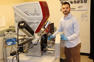 It is incredibly frustrating and seemingly paradoxical how quickly soft materials, such as hair, dull razor blades and other hard materials. Gianluca set out to explain this behavior through in-situ SEM. As he learned in his studies, there are three major factors that contribute to hair's ability to fracture steel: (1) the angle at which the hair approaches the blade, (2) the relative position of hair to features on the blade, and (3) the intrinsic heterogeneity of the steel's martensite structure. It is incredibly frustrating and seemingly paradoxical how quickly soft materials, such as hair, dull razor blades and other hard materials. Gianluca set out to explain this behavior through in-situ SEM. As he learned in his studies, there are three major factors that contribute to hair's ability to fracture steel: (1) the angle at which the hair approaches the blade, (2) the relative position of hair to features on the blade, and (3) the intrinsic heterogeneity of the steel's martensite structure.
Only the third factor, the heterogeneity of the material, is engineerable, so Gianluca worked to design better blades with this information in mind. He has been very successful in this pursuit and is launching a start-up after his imminent graduation called RazorSharp with his patented technology. Check his website to learn more about this!
Gianluca's passion for electron microscopy is self-evident, and as he states more explicitly, he loves that microscopy gives him the ability to better understand and explain things.
When he's not in lab, Gianluca is out walking, relaxing at the beach, cooking, developing his photography skills, and looking after his "happy fish" in his 55-gallon tank. As he points out, the same principles apply to microscopy and photography, making this an apt hobby for someone in this field.
A team of scientists at Northwestern led by Dr. Linus Segbauer published a paper in PNAS describing the complex structure of the rock-grazing mollusk Cryptochiton stelleri tooth. Nano-disperse, amorphous iron hydroxyphosphate is present in the stylus, extending the range over which hardness and stiffness vary by at least a factor of two. This discovery was made through a number of correlative microscopy and spectroscopy techniques, including work at Argonne's APS. The significance of this work extends beyonds its biological implications as this understanding could help with the design of new kinds of materials by creating chiton-inspired ink for 3-D printers.
Region VII Update:
Research performed at Rice University has shed light on the unique toughness of hexagonal boron nitride. This project combined experimental microscopy performed in the labs at Rice and theoretical work performed at Nanyang Technological University in Singapore to probe the crack propagation mechanisms that give hBN its unique toughness and how it differs from the observed brittle behavior in graphene. This fracture-resistant behavior in hBN opens it up as a very useful material for 2D and flexible electronics. Rice Press Release Article
Like what you see? Join our Social Media Committee
- Help MSA students create informative, engaging content for microscopists and the greater public
- Responsibilities tailored to your schedule, varying from Instagram content creation to outreach and engagement during M&M
- Reach out for more info: studentcouncil@microscopy.org
|
|
Local Affiliated Societies
|
|
Local Affiliated Societies News
by Ru-ching Hsia, LAS Director
MSA members across the country have also established 25 local affiliated societies (LAS). These LAS hold regular meetings, social events and publish newsletters to provide regional networking and outreach opportunities for the local microscopy community.
Regional LAS activities
The 56th annual meeting for the Southeastern Microscopy Society' (SEMS) will be held virtually on June 24, 2021! Please visit the SEMS website (http://southeasternmicroscopy.org/meetings/) for meeting Flyer, Agenda and the "Call for Papers" information for the Ruska Student and the Robert Simmons Micrograph Competitions.
LAS Programs
LAS Business Meetings
LAS Business Meeting of 2021 will be held on the second Thursday in the month of March, June, September, and December. Officers and Members of LAS are welcome to attend the meeting. Different LAS will be featured in each meeting and present their activities and events. We will also share ideas and tips for running local societies and events during the meeting. LAS are encouraged to work with regional liaisons from the MSA Student Council (StC), local colleges, and other regional scientific or teachers' societies, etc.
The next LAS business meeting will be held on June 10th, 1 to 2 PM EDT. Arizona Imaging and Microanalysis Society (AIMS) and Oklahoma Microscopy Society (OMS) will share their experiences on their most recent virtual meetings. Treva Brown, the Director of the Diversity, Equity, and Inclusion (DEI) committee of the MSA will also be present to give us an introduction on many initiatives the DEI committee have been implementing.
If you are interested to start an Affiliated Society in your region or have any questions and concerns, please feel free to contact me at rhsia@umaryland.edu.
Thanks!
Ru-ching
|
|
|
|
|
Biomedical Microscopy Focused Interest Group (DBM-FIG) will be hosting a series of "BioEM Talks" on four consecutive Fridays on July 31st, August 6th, 13th and 20th from 10:00 AM to 1 PM EDT (2 to 5 PM GMT). This series is to replace the Pre-meeting congress (PMCX 61) that was supposed to be held on the Sunday before the M&M this year. Each session in the series will include live presentations of three invited speakers, vendor exhibits and pre-recorded flash talks in the Gather Town virtual space. Attendees are encouraged to submit 5 min video flash talks along with a PowerPoint presentation of no more than ten slides. These flash talks (replacing traditional poster boards) will be grouped and presented on demand in the BioEM virtual Town Hall along with vendor exhibits. Tentative program and registration information can be found at the DBM-FIG website ( https://diagnosticbiologicalmicroscopy.com/) .
BioEM Talks are free and open to all to attend. We hope this series will serve as a new platform to stimulate more collaboration nationally and internationally in the biological EM community. Please feel free to download the BioEM Talks flyer and help us spread the word. Contact the chair of the organizing committee, Dr. Ru-ching Hsia ( rhsia@umaryland.edu) for any inquiries.
|
|