Looking for a specific section? Find it quick with these links below!
|
 |
|
There's Still Time to Renew Your MSA Membership for 2021
If you haven't already, time is running out to renew your membership to avoid a lapse in member benefits at the end of the month! Renew your membership today and remain part of the community that brings microscopy and microanalysis professionals of all levels together.
|
|
If you are interested in learning more or joining the task force, please reach out to Dr. Brown at [email protected].
|
|
Spotlight on MSA's Diversity, Equity, and Inclusion Task Force
Last year's annual meeting featured a three-part "Lens on Diversity" series co-led by Dr. Treva Brown, MSA member and Chair of MSA's Diversity, Equity, and Inclusion Task Force. Hear from Dr. Brown as she discusses the importance of diversity and representation in the physical, biological, analytical sciences, and within the Society. |
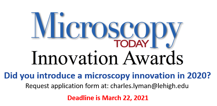
Submit an Article to Microscopy Today
The Editors of Microscopy Today (MTO) encourage and greatly appreciate submission of articles from microscope users as well as microscope manufacturers and suppliers. Of particular interest are summaries of in-depth articles published in peer reviewed journals and articles that describe new equipment and applications. Microscopy Today is open access and there are no charges for publishing in MTO. All articles are available free to our subscription list of over 18,000 microscopists and through our collaboration with Cambridge University Press over 8,000 libraries worldwide. For further information email the Editor-in-Chief at [email protected] or visit
|
|
Upcoming Microscopy Today Webinar
|
Ultrafast Transmission Electron Microscopy: Techniques and Applications Tuesday, May 11, 2021 | 12:00 p.m. ET
With the growing applications of temporally-resolved electron microscopy for basic phenomena and reducing damage in samples, this webinar will provide an introduction to the field of ultrafast transmission electron microscopy, including techniques, equipment and historical perspectives from technical experts. A combination of historical developments with recent advances will help attendees in considering ultrafast capabilities for their own research, or satisfy general interests. The ultrafast technology review will also include a broader scope of applications enabled by ultrafast techniques using a variety of sample stimuli from multidisciplinary fields. Register here.
|
|
|
Abstract submission is now open for Microscience Microscopy Congress (mmc) 2021
Taking place virtually for the first time ever!
While many public events around the globe continue to be postponed or cancelled due to COVID-19, the RMS has risen to the challenge of running mmc2021 as a one-hundred-per-cent, virtual conference and exhibition - the biggest online event of its kind in Europe - from 5 - 9 July.
The international event combines six parallel streams of conference sessions with a free-to-attend exhibition, plus virtual meetings and workshops covering all the latest topics. It serves as a golden opportunity for companies - both large and small - to showcase a quantity and quality of equipment that never fails to impress. It also provides the perfect stage for delegates to showcase their work, make new connections and broaden their knowledge base and skill-set.
Abstract submissions, in the form of both oral and poster presentations, are now open at www.mmc-series.org.uk/abstract-submission. All abstracts should be submitted through the dedicated online system, which is being hosted by Oxford Abstracts. The deadline is 18 March 2021.
To find out more about the biggest virtual event of its kind in Europe, and submit your abstract, visit www.mmc-series.org.uk.
|
|

|
 The MSA Facebook page regularly posts science news for you The MSA Facebook page regularly posts science news for you
New AI tool can revolutionize microscopyAn AI tool developed at the University of Gothenburg offers new opportunities for analyzing images taken with microscopes. A study shows that the tool, which has already received international recognition, can fundamentally change microscopy and pave the way for new discoveries and areas of use within both research and industry. Read more.

|
Women's History Month: 50+ Women in Science and Engineering to Learn More About
There have always been women in science. Whether recognized at the time or not, women interested in science and engineering have made important discoveries, conducted game-changing research, and pursued careers in science, technology, engineering, and math (STEM). Uncovering and sharing these stories and encouraging girls to see themselves as future scientists is important, ongoing work for science educators. Read more.
|
|
|
|
|
|
MSA Student Council Executive Council
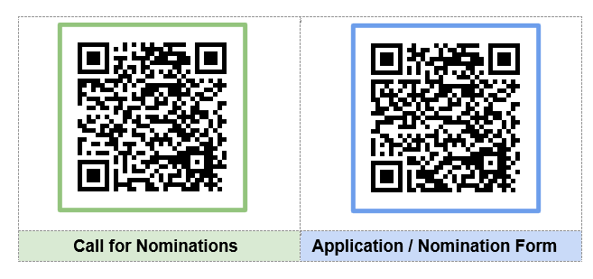
Call for Nominations opens March 15th!
The Call for Nominations for StC elected positions opens March 15th
and will close on April 30th.
A description of the roles for the three executive positions (President-Elect, Secretary, and Treasurer) can be found on the MSA Student Council website, Responsibilities page.
- President-Elect serves a three-year term, one each as President-Elect, President, and Past-President
- Secretary and Treasurer positions serve a one-year term.
Graduate and undergraduate students that are members of MSA are encouraged to apply. This is a wonderful opportunity to hone your professional skills in leadership, communication, conference planning, and networking while advancing the growth of the student community in MSA.
The Student Council cordially invites all MSA undergraduates, graduate students, and postdocs to attend our 2nd Student and Postdoc social - a Virtual Game Night on Tuesday, March 30th from 8:00-9:30 pm Eastern Time! Game options will include Among Us, Codenames, and Scattegories. All attendees will be entered into a raffle to win a $25 Amazon gift card! Please RSVP using this link by Friday, March 19th for planning purposes and to receive instructions for accessing the Discord server.
Missed Microscopy Live?
Check out the recording of our last Microscopy Live webinar, featuring Prof. Wenpei Gao's live demonstration of 4D-STEM: https://youtu.be/ZF6DARp2cCs
Matthew Cheng - Physical Sciences
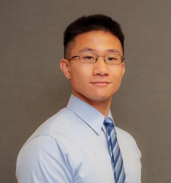 Congratulations to Matthew Cheng on being MSA StC's March Physical Sciences Student Spotlight! Congratulations to Matthew Cheng on being MSA StC's March Physical Sciences Student Spotlight! Meet Matt: Matt Cheng is a 4th year PhD student at Northwestern University in the group of Professor Vinayak Dravid. Matt graduated from University of Illinois Urban-Champaign (UIUC) in 2016 from the Department of Materials Science and Engineering. Research Focus: Matt's area of expertise is on characterizing 2D materials with an emphasis on high resolution electron microscopy. He works to establish structure-property relationships in 2D ferroic materials for next generation memory storage and quantum devices. This ties into larger efforts in the field of device development to miniaturize new technologies. Road to graduate school: Matt was sold on the idea of going to graduate school by a formative research experience as an undergraduate at UIUC. In particular he cites the mentorship of an at the time graduate student, Dr. Priya Raman, who helped him get started in lab during his first research experience. The PhD experience: Matt is enjoying his PhD, and the opportunity to study materials on the atomic/molecular level. In particular, he likes working in the Dravid group because he is able to take ownership of his own project, while collaborating with teams in other groups and at Northwestern's NUANCE center.
Why microscopy? As Matt explains, electron microscopy is an incredible tool to be able to observe fundamental properties of materials and develop an understanding of their behavior. He especially loves the high-resolution electron microscopy and being able to see the organization of atoms in a material. However, he also mentions that these exhilarating research discoveries are sparsely spread out by the real challenge - properly preparing a sample for analysis, a time consuming and sometimes tedious process. Nonetheless, Matt loves microscopy and the contributions it allows him to make in the study of 2D materials.
TAing (during a pandemic!): Recently, Matt has taken on the challenge head-on of TAing an introductory graduate course on electron microscopy. Given COVID restrictions, the classes are virtual, so there is no lab component of the course. Nonetheless, he's been doing a fantastic job connecting with students and helping them learn in different ways given their individual constraints. He describes the experience by saying - "as a student you're supposed to learn quite a bit, but as a TA you learn even more."
Advice to those getting started in the field? Don't be afraid to ask questions and fail fast.
Beyond science: When Matt is not in lab, he loves to cook - although he points out that cooking is somewhere between art and science anyways. He is great at BBQing but has recently been doing a lot of baking. He is also an avid hiker, but will settle for a trip to the gym, given the limited outdoors space in Chicago.

Kelly Parker - Biological Sciences
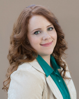 The March Biological Sciences Student Spotlight is a fifth year PhD student at Northwestern University in Dr. Vinayak Dravid's research lab. Her research involves characterizing soft and biological materials, specifically proteins, DNA, and other small biological molecules. Because these materials are both low-contrast and susceptible to beam damage when imaging, Kelly's research focuses on improving contrast while avoiding high electron doses; this includes using low-voltages, particularly in scanning electron microscopes with transmission detection (STEM), for high-contrast, high-throughput imaging. Kelly's work, thus, enhances the imaging tools required to answer questions related to the characterization of various biological materials. The March Biological Sciences Student Spotlight is a fifth year PhD student at Northwestern University in Dr. Vinayak Dravid's research lab. Her research involves characterizing soft and biological materials, specifically proteins, DNA, and other small biological molecules. Because these materials are both low-contrast and susceptible to beam damage when imaging, Kelly's research focuses on improving contrast while avoiding high electron doses; this includes using low-voltages, particularly in scanning electron microscopes with transmission detection (STEM), for high-contrast, high-throughput imaging. Kelly's work, thus, enhances the imaging tools required to answer questions related to the characterization of various biological materials.
Kelly completed her undergraduate studies at Stanford University with a degree in Materials Science and Engineering. While at Stanford, she completed a summer internship at the Leibniz Institute for New Materials in Germany. She is in a unique position as a material scientist studying biological processes. She is particularly interested in biotechnology and medical device research, and plans to pursue a career in this area.
Kelly has been to three Microscopy and Microanalysis (M&M) meetings, including 2020's virtual program, and wishes she could combine aspects of both for the ideal conference. During the virtual format, for example, she was able to find the talks she was interested in and view them on her own time - this was especially true for presentations that occurred simultaneously. She also enjoyed the multiple diversity discussions. She did, however, miss seeing and meeting attendees in person.
As a woman in STEM, Kelly is especially vested in diversity and inclusion programs. She is currently in her third year as the Mentorship and Outreach Chair of the Graduate Society of Women Engineers (GradSWE) at Northwestern. Kelly leads a committee of three other women to help put on community outreach events and manage mentorship programs within the university. Her focus, she says, is "supporting girls and other underrepresented minorities" in her local community.
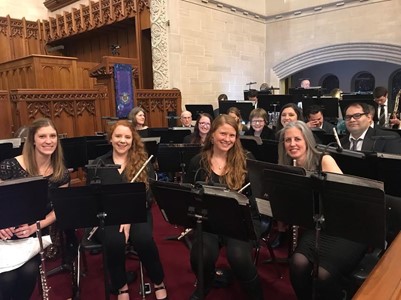 Outside the lab, Kelly is a self-proclaimed nerd and we love it! She loves playing Dungeons & Dragons (D&D) - she is in two groups, one of which was born out the pandemic. She is also a classical musician - playing the flute for fifteen years - and is a member of the Windy City Winds, a community wind ensemble, where she plays the piccolo. Kelly and her friends have also formed a group called Snarky Winds and have been making virtual concert videos. Find them on YouTube! Outside the lab, Kelly is a self-proclaimed nerd and we love it! She loves playing Dungeons & Dragons (D&D) - she is in two groups, one of which was born out the pandemic. She is also a classical musician - playing the flute for fifteen years - and is a member of the Windy City Winds, a community wind ensemble, where she plays the piccolo. Kelly and her friends have also formed a group called Snarky Winds and have been making virtual concert videos. Find them on YouTube!
We are thrilled that Kelly is leading the way for young future scientists and are excited for her own future endeavors, both scientific and social.
Region II Update
Happy women's history month! On International Women's Day (March 8th), the NUANCE center and MSA student council teamed-up to host a virtual conference celebrating women in microscopy. The event kicked-off with a welcome from MSA-president elect Prof. Deb Kelly and NUANCE center director, Prof. Vinayak Dravid, followed by a keynote presentation from Dr. Ilke Arslan. The rest of the day was comprised of research talks in a variety of areas including cryo-EM, magnetism, and quantum imaging. The conference concluded with a series of career panels from vendors, lab managers and researchers in both academic and federal labs. Stay tuned - all conference talks will be posted on YouTube for later viewing in case you missed the conference.
Region V Update
An interesting new paper has published where researchers have been able to image the crystallographic 32 Å moiré pattern at the 2D/3D interface using scanning TEM. This work provides a general technique on how to directly image periodic modulation at interfaces by combining various emerging new electron microscopy techniques. The research was published in Nature Communications - Reidy et al. "Direct imaging and electronic structure modulation of moiré superlattices at the 2D/3D interface", DOI: http://doi.org/10.1038/s41467-021-21363-5
Region VIII Update
The Tillberg Lab at Janelia Research campus, in conjunction with Utrecht University recently published a paper putting forth a novel sample preparation technique for bypassing the diffraction limit in light microscopy. Their Ten-fold Robust Expansion Microscopy (TREx) provides an improvement to the Expansion Microscopy technique without the requirement of specialized equipment and procedures for the sample preparation. Congrats to the team over at Janelia Research Campus. Janelia Abstract
Like what you see? Join our Social Media Committee
- Help MSA students create informative, engaging content for microscopists and the greater public
- Responsibilities tailored to your schedule, varying from Instagram content creation to outreach and engagement during M&M
- Reach out for more info: [email protected]
|
|
Local Affiliated Societies
|
|
Local Affiliated Societies News
by Ru-ching Hsia, LAS Director
MSA members across the country have also established 25 local affiliated societies (LAS). These LAS hold regular meetings, social events and publish newsletters to provide regional networking and outreach opportunities for the local microscopy community.
Regional LAS activities
Arizona Imaging and Microanalysis Society (AIMS) will be hosting a one day virtual conference on March 19, 2021 and an ImageJ/Fiji Image Analysis Workshop on March 18, 2021. Check out the event information at http://azmicroscopy.org/events/.
Oklahoma Microscopy Society (OMS) will be hosting a virtual Spring meeting on April 22-23, 2021. Agenda includes scientific research presentation, new product introduction and image analysis workshop. Free to attend, Register at https://okmicroscopy.org/.
LAS Programs
LAS Business Meetings
LAS Business Meeting of 2021 will be held on the second Thursday in the month of March, June, September and December. Officers and Members of LAS are welcome to attend the meeting. Different LAS will be featured in each meeting and present their activities and events. We will also share ideas and tips for running local societies and events during the meeting. LAS are encouraged to work with regional liaisons from the MSA Student Council (StC), local colleges, and other regional scientific or teachers' societies, etc.
If you are interested to start an Affiliated Society in your region or have any questions and concerns, please feel free to contact me at [email protected].
Thanks!
Ru-ching
The AIMS meeting is scheduled for Friday, March 19, 2021 using a virtual platform. The conference link is AIMS2021.vfairs.com.
The ImageJ/FIJI workshop is scheduled for Thursday, March 18, 2021 and will be presented as an online TEC Talk workshop. For more information and to register, click here.
|
|
|
|
|
|
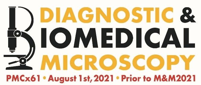
Missed the M&M abstract deadline? There is still time to submit your abstract to the Pre-Meeting Congress X61.
|
Renu Sharma, Chair
PMC X61 is a full day symposium organized by Diagnostic and Biomedical Microscopy Focused Interest Group (DBM-FIG). It will be held on the Sunday of August 1st and focused entirely on the biological electron microcopy research and technologies. Bruno Humbel of Okinawa Institute of Science & Technology and Kristina Micheva of Stanford University will be keynote speakers. The same abstract submitted to the M&M symposia can be submitted to the PMC as well. Please go to https://diagnosticbiologicalmicroscopy.com/pmc-x61/ for more information about PMC X61 and submit abstract ( https://diagnosticbiologicalmicroscopy.com/attend/). The PMC X61 abstract submission deadline has been extended to Friday, April 16, 2021, 11:59 PM, EST.
|
|