Looking for a specific section? Find it quick with these links below!
|
|
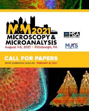
Look for the M&M 2021 Call for Papers Brochure with the November/December issue of Microscopy Today. For more information on M&M 2021, visit the website at https://www.microscopy.org/MandM/2021/
The Call for Papers submission site will open December 7, 2020. All papers must be submitted by February 18, 2021.
|
|
 |
Renew Your MSA Membership for 2021!
The membership renewal process is currently underway! Keep your membership current to maintain access to exclusive member benefits. The deadline to renew is December 31, 2020. Take it a step further and consider adding a donation when you renew to help sustain the Society as we head into 2021. We hope that you'll take a moment to renew your membership and remain part of the community that brings microscopy and microanalysis professionals of all levels together. Renew today!
Upcoming Microscopy Today Webinar sponsored by 3D Micromac
Preparation of a sample can take a while and is often challenging. In times when the production of materials and products becomes more and more complex, and components are getting smaller, also test, analysis, and diagnostics methods are becoming more complex and expensive. There will be a high need to speed up these processes, simplify them in advance and tailor them to the appropriate procedures. Register here.
|
|

|
|
 The MSA Facebook page regularly posts science news for you The MSA Facebook page regularly posts science news for you
Cryo-electron microscopy reaches atomic resolutionA founding principle of structural biology is that, once researchers can directly observe macromolecules in enough detail, it should be possible to understand how their 3D structures confer their biological functions. Indeed, many scientific advances have relied on directly observing the world around us in as much detail as possible, and efforts are increasingly being dedicated to visualizing the atomic structures of biological components that have a key role in human disease. Nuclear magnetic resonance (NMR) spectroscopy, X-ray crystallography and cryo-electron microscopy (cryo-EM) are the three main structural-biology techniques in use. Of the three, cryo-EM has emerged as the current 'go to' method for determining the structures of large and dynamic complexes that have proved difficult to obtain by the other approaches. Read more.
World record resolution in cryo electron microscopy
Holger Stark from the Max Planck Institute for Biophysical Chemistry in Göttingen and his team have broken a crucial resolution barrier in cryo electron microscopy. For the first time, his group succeeded in observing individual atoms in a protein structure and taking the sharpest images ever with this method. Such detailed insights make it easier to understand how proteins do their work or cause diseases in the living cell. The technique can also be used in the future to develop new drugs. Read more.
|
|
|
|
|
Student Council is continuing our tradition of holding educational and informative webinars with our series this year on the following topics:
- Proposal Writing and Applying for Funding
- Diversity and Inclusion in Microscopy
- The Undergraduate Experience
- Ask a Microscopist
- Communicating Science: Data Visualization
- Microscopy Careers Mini-series (Professors, Industry, National Labs)
- Preparing for M&M
StC is excited to announce that any MSA member who attends all webinars in this series will be entered in a drawing to win a StC merchandise package including a T-shirt, mug and more!
Are you a PI, Technician, Post-doc, or other microscopist that is experienced or knowledgeable in any of the above topics? We'd love to have you as a panelist in one of these webinars! Please fill out the form at the following link to inform us of your interest: https://forms.gle/Sz9dQtutuVqVGgTX6
Social Media Committee Is Now Recruiting
- Looking to get involved? Help us create content for microscopists (informational) and for public outreach (creative)
- Responsibilities tailored to your schedule
- Contact Chris or Louisa for more information: [email protected]

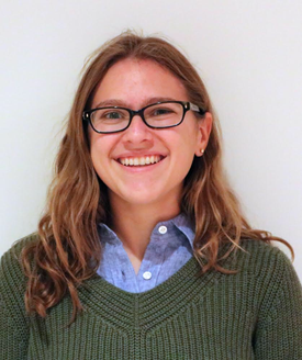
Juan Sanchez - Biological Sciences
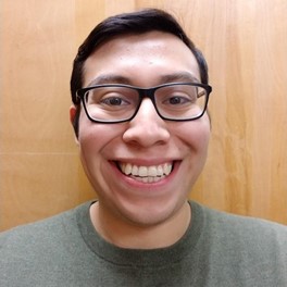 November's Biological Sciences Spotlight is Juan Sanchez, a second-year biophysics student in Dr. Elizabeth Wright's lab where he studies how bacteriophage interact with prokaryotic appendages. He uses cryo-EM to analyze the structure of flagella and pili to understand how they function in an effort to better develop antimicrobials to provide new treatments for infectious diseases. Juan presented his research at his first attendance to the Microscopy and Microanalysis (M&M) Conference this year, where he described producing single protein caulobacter flagella in order to understand the function of each of the six proteins that comprise each flagella.
In addition to the scientific talks Juan attended at this year's M&M, he mentioned that the Diversity and Inclusion Lens On seminars were a highlight of the week, saying that he felt a sense of community outside the science that initially brought the attendees to the meeting. He adds, "I really felt a part of the meeting."
Juan completed his undergraduate degree in biology at Virginia Commonwealth University. However, with no concrete plan laid out following graduation, he moved to Chicago and found a job as laboratory technician at a nearby college. But as luck would have it, he met someone who was pivotal in his path to his graduate studies. He completed his master's degree while working full time and doing research in the lab where he would ultimately wind up for his doctoral studies.
Juan's interests after completing his PhD involve pursuing a position in research and development. As part of his efforts to reach that goal, he applied for and was awarded a T32 grant through the University of Wisconsin-Madison, where he now studies. With this biotechnology training fellowship, Juan will be working to help bring a patent idea to market, and eventually have the opportunity to explore various industry internships.
 His experiences as a first-generation college student along with the seemingly unconventional path he took to graduate school has made Juan realize that there might be other students out there unsure about the possible fields of study available. To this end, he is passionate about exposing young high school students to careers other than the obvious doctor or lawyer. He was awarded a SciMed GRS fellowship through his university to do just that. "You only know what you know", he says, encouraging students to explore what's out there. He also urges new graduate students, especially minority students, to find a community that you feel comfortable in. He explains that it's "sometimes overwhelming being the only minority in the room" and that having a group of people who can relate to you alleviates much of the pressures of graduate school.
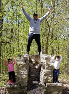
Any free time Juan has away from the lab is spent with his family. He and his wife enjoy playing recreational soccer and he even volunteered to coach his 4-year-old son's soccer team, which he amusingly called "a mess". His son and 3-year-old daughter, he says, "are my greatest motivation to be productive in the lab".
Emily Greenstein - Physical Sciences
 Meet November's Physical Sciences student spotlight, Emily Greenstein. Emily is a 3rd year PhD student in the Materials Science and Engineering department at Northwestern University. Emily has diverse talents, which enables her to work on all aspects of her project: from theory to synthesis to microscopy. This is in part facilitated by the fact that she is advised by both Professor Ken Poeppelmeier (Chemistry) and Laurence Marks (Materials Science). Emily researches heterogeneous catalysis and nanoparticle synthesis, working on optimizing catalysis design for improved performance in a variety of applications, including CO conversion. The nanoparticles, which serve as catalytic supports, are grown with a novel "hydro-sauna" or vapor assisted sol gel approach. She then images the interfaces of these catalysts with high resolution TEM to understand how the surfaces of these materials impact their performance. Meet November's Physical Sciences student spotlight, Emily Greenstein. Emily is a 3rd year PhD student in the Materials Science and Engineering department at Northwestern University. Emily has diverse talents, which enables her to work on all aspects of her project: from theory to synthesis to microscopy. This is in part facilitated by the fact that she is advised by both Professor Ken Poeppelmeier (Chemistry) and Laurence Marks (Materials Science). Emily researches heterogeneous catalysis and nanoparticle synthesis, working on optimizing catalysis design for improved performance in a variety of applications, including CO conversion. The nanoparticles, which serve as catalytic supports, are grown with a novel "hydro-sauna" or vapor assisted sol gel approach. She then images the interfaces of these catalysts with high resolution TEM to understand how the surfaces of these materials impact their performance.
Emily has incredibly broad enthusiasm for science, which extends even beyond the scope of her project. For example, she has recently been working on building a new synthesis reactor, which involves the challenge of negotiating the world of piping, tubing, and valves. Although this work falls far outside the scope of heterogeneous catalysis, Emily was excited about this unexpected aspect of her project, and even mentioned that it had some side benefits in recent home repairs. She has been involved in mentorship through the National Student Leadership Conference, where high school aged students live in a college setting to meet scientists and continues to serve through Expanding Your Horizons, a conference for girls to meet STEM female role models. Emily described in her experience the best mentors facilitate joy of learning through enthusiasm about the material they're teaching and openness to questions. Emily certainly embodies this herself! When asked to provide advice for new researchers, she suggests not to get too bogged down in details and to learn by doing.
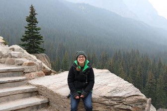 Outside of the lab, Emily loves puzzles, outdoors, and wildlife photos. In particular, she likes to take photos of birds. Emily described the relationship between microscopy and birding saying that both require patience, pattern recognition and an appreciation for the sensitivity of your material. It is our pleasure to have Emily as our November student spotlight and look forward to hearing more about her research (and birding) as her graduate school career progresses. Outside of the lab, Emily loves puzzles, outdoors, and wildlife photos. In particular, she likes to take photos of birds. Emily described the relationship between microscopy and birding saying that both require patience, pattern recognition and an appreciation for the sensitivity of your material. It is our pleasure to have Emily as our November student spotlight and look forward to hearing more about her research (and birding) as her graduate school career progresses.
We're excited to report that University Wisconsin has received a $22.7 million 6 year grant from the NIH to create a national center for Cryo-EM. Madison will serve as the central hub that includes Stanford, University of Colorado-Boulder and the New York Structural Biology Center. One of the main focuses of the center is on tomography (Cryo-ET), a three dimensional imaging technique, with an emphasis on biological molecules. One application of Cryo-ET is in drug discovery, as the imaging technique can help understand the interaction of diseases and potential therapies. This is an incredibly exciting announcement for the biological microscopy community at large, but especially in the Midwest. Congratulations to the team at Wisconsin!
Optical Microscopy has been used to study complex biological systems but is often limited by the time taken for 3-D volumetric data acquisition. Recently, researchers from Biomicroscopy Lab at the Boston University have developed a simple and versatile system to tackle this problem by developing a novel image acquisition technique. This technique enables to capture the wide field image stack using a single camera and record it simultaneously. This new system can be readily applied to fluorescence, phase-contrast and dark field imaging. The research article was published in OPTICA - S. Xiao, H. Gritton, H. Tseng, D. Zemel, X. Han, and J. Mertz, "High-contrast multifocus microscopy with a single camera and z-splitter prism," Optica 7, 1477-1486 (2020).
Oak Ridge National Lab has published their report on the atomic structure of the Protease enzyme of Covid-19. This data contains key information about replication mechanisms and the data set has been made fully available to facilitate research around the globe. A process that can often take years for a protein of this size was completed and published in just 5 months. Congratulations to the neutron scattering team at ORNL! For further information, as well as the published article: ORNL Press Release.
|
|
Local Affiliated Societies
|
Local Affiliated Societies News
by Patty Jansma, LAS Director
LAS Meetings
Check the individual LAS websites for more details.
Support your local affiliated society! Invite students, early career scientists and technologists to your LAS meetings. Better yet, bring a new member to your local meeting and get them involved!
LAS Programs
As always, you may contact me at [email protected] with comments, questions or concerns.
|
We're excited to announce the new LAS Director elected to MSA Council for 2021-2023!
Join us in welcoming Ru-Ching Hsia as the newly-elected LAS Director. We look forward to the new things in store beginning next year!
|
|
|
|
|
|
|
Renu Sharma, Chair
Join a FIG! FIGs are groups of scientists that practice or have interests in specific disciplines (currently 11) to which microscopy and microanalysis is applied. As an MSA member, you can join one or more (FIG Communities). FIGs not only boost scientific understanding through knowledge sharing, but also provide opportunity to network with scientists who share common interests. FIGs may organize lunches, symposia or pre-meeting congresses at M&M. A complete list of FIGs is on MSA website or by clicking here. You may contact the FIG leader directly or attend a business meeting at M&M to learn more. Visit the FIG Store to sign up. Are you already a member of a FIG? Consider volunteering and make an impact! It's members are what makes FIGs successful. Talk to your FIG leader. Interested in starting a FIG? Start by reviewing the updated version of the FIG Guidelines and then contact me. FIGs are for students too! If you are a student, your fees are waived for the first FIG you join.
Are you interested in highlighting your FIG in an MSA Update? Contact me for more information.
|
|