CFS Logo

Retinoblastoma Foundation

Centre of Excellence in Ocular Oncology

Centre for Sight Super-specialty Eye Hospital

Banjara Hills, Hyderabad, India


Retinoblastoma - They Live and See

Optimizing Life, Eye and Vision Salvage


A world retinoblastoma awareness week initiative


Part 1. Symptoms, Signs, Diagnosis

How common is retinoblastoma?


Retinoblastoma is the most common malignant intraocular tumor in children. Incidence is variable, and is estimated to occur in 1 in 10,000 to 15,000 live births. India leads in the annual incidence of retinoblastoma with about 1600 - 2000 new cases every year.


At what age does retinoblastoma manifest?


A majority of cases of retinoblastoma present to an ophthalmologist between 9 months to 36 months of age.


Is retinoblastoma unilateral or bilateral?


About 60-70% of retinoblastoma are unilateral and unifocal (most likely sporadic), while 30-40% are unilateral and multifocal or bilateral (heritable).


Is genetic test important in a child with retinoblastoma?


Every child with retinoblastoma and the parents are ideally subjected to an appropriate genetic test to assess prognosis, triage the frequency of screening of the contralateral eye, need and frequency of screening of the siblings, and determine the risk of retinoblastoma in the family.

What are the most common symptoms of retinoblastoma?


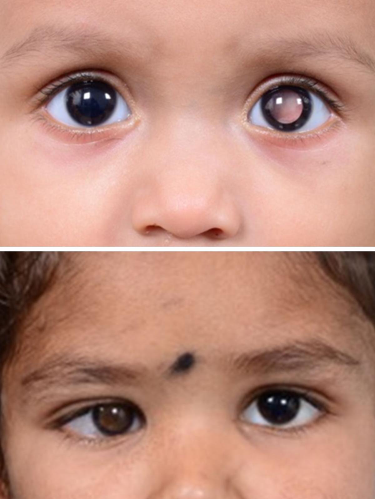
White reflex (50%) and strabismus (25%) are the two most common symptoms as well as signs of retinoblastoma. Creating parental awareness that any child with an abnormal reflex or squint needs to be immediately shown to an ophthalmologist can help in early diagnosis.


Pediatricians and comprehensive ophthalmologists must be taught to screen for a white reflex in every child that they see. Ophthalmologists must be aware of the sinister significance of white reflex and squint in a child and must examine these children thoroughly and in case of doubt, expeditiously refer to a retinoblastoma treatment centre.


White reflex (top) is the presenting symptom and sign in 50-60% and squint (bottom) in 15-25% children with retinoblastoma

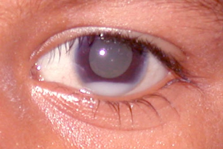
Tumor deposits in the anterior chamber simulating hypopyon in a 5-year-old child with retinoblastoma

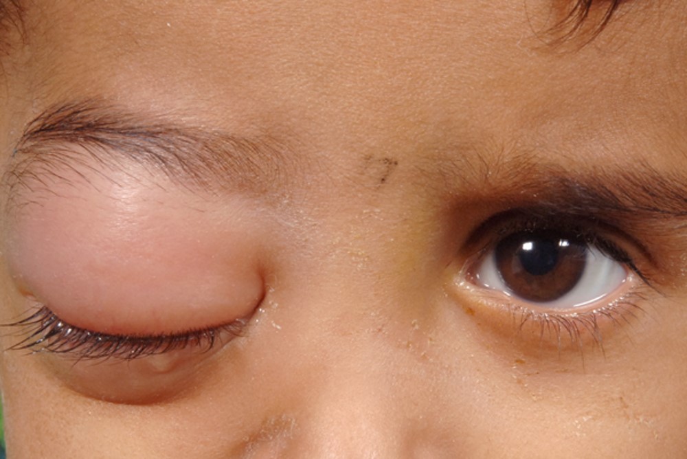
Sterile orbital celllulitis secondary to inflammation induced by spontaneous tumor necrosis in a 2-year-old child with retinoblastoma

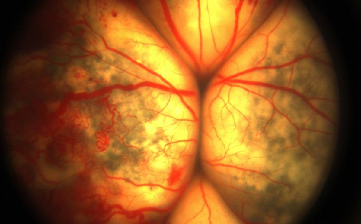
Golden yellow reflex is typical of Coats' Disease. Not characteristic peripheral retinal telangiectasia, extensive exudation and exudative retinal detachment.

What are the masquerades of retinoblastoma?


While masquerades such as hypopyon, hyphema, uveitis, vitritis, vitreous hemorrhage and retinal detachment are more common in an older child with retinoblastoma, infants do present with buphthalmos, cataract, orbital cellulitis as a consequence of tumor necrosis and sterile inflammation and phthisis of unexplained etiology.


A high index of suspicion and appropriate investigations are necessary to rule out retinoblastoma in such situations, especially if an invasive intraocular procedure is considered.


What are the differential diagnosis of retinoblastoma?


Five common causes for leucocoria simulating retinoblastoma are - Coats' Disease (xanthocoria, peripheral retinal telangiectasia, exudates), PHPV (relative microphthalmos, prominent ciliary processes), Retinal Astrocytoma (transluscent retinal lesion, associated tuberous sclerosis), Toxocara (tractional elements - falciform fold and dragged-disc appearance) and Medulloepithelioma (anterior ciliary body location, zonular coloboma).

How is retinoblastoma diagnosed?


A comprehensive examination of both the eyes under anesthesia is essential to diagnose retinoblastoma and determine clinical risk factors to triage baseline investigations.


Presence of clinical risk factors warrants baseline bone marrow biopsy and cerebrospinal fluid cytology. A thorough indented evaluation of the retinal periphery is essential to screen for retinoblastoma in the contralateral eye if the disease is apparently unilateral.

How is retinoblastoma staged?


Retinoblastoma is prognosticated by categorizing it into Groups and Stages. While eye salvage is the outcome measure for grouping, staging has implication on life salvage.


International Grouping of Intraocular Retinoblastoma has five groups - A to E. International Retinoblastoma Staging System has Stage 0 to IV. The new AJCC 8 TNM classification additionally takes heritability into consideration.

Does imaging have a role in the diagnosis and management of retinoblastoma?


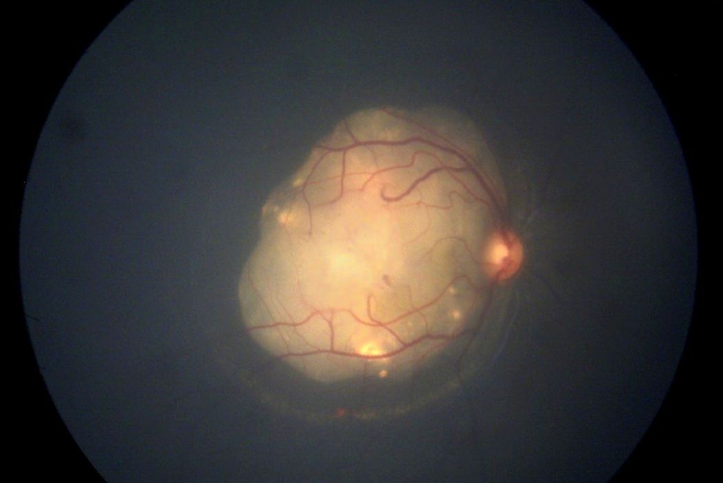
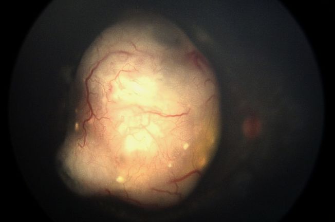
  1. Baseline and serial wide-field fundus photography is essential for documentation of the extent of the tumor and its resolution and to detect early recurrence
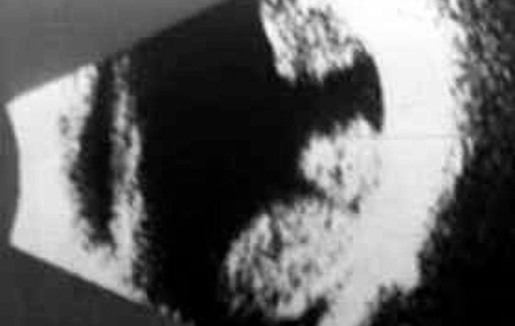
  2. Ultrasound B-scan helps in supporting the clinical diagnosis, localize a tumor if the tumor is exophytic or if the media is hazy, and measure the tumor.
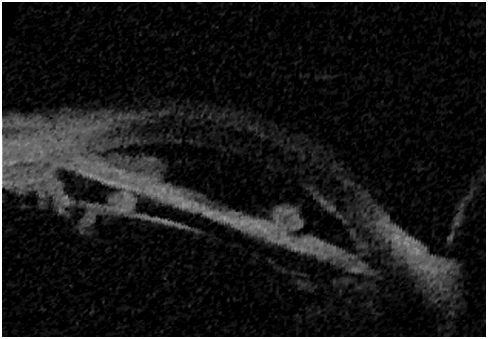
  3. Ultrasound biomicroscopy is essential to evaluate the presence and extent of ciliary body invasion if clinically suspected.
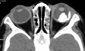
  4. CT scan helps detect intraocular calcification as a supporting diagnostic sign in confounding situations and can be a useful adjunct but is ideally avoided because of risk of radiation exposure.
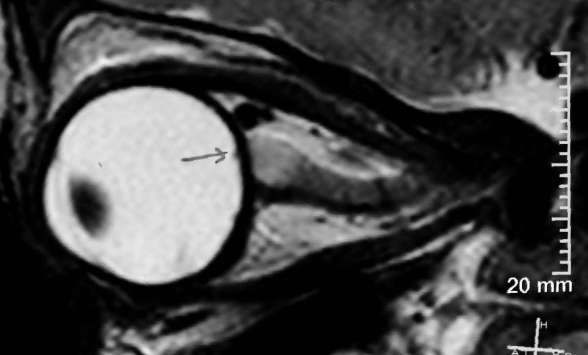
  5. A baseline MRI is essential in bilateral retinoblastoma to screen for pineloblastoma, and in Group D and E tumors to evaluate for choroidal invasion, optic nerve infiltration and extraocular extension.
  6. PET-CT scan is useful in screening macroscopic systemic metastasis when indicated.

Wide-field RetCam fundus images are extremely useful for baseline documentation and for monitoring treatment response.

Ultrasound B-scan helps detect intraocular calcification, and also measure the tumor


Ultrasound biomicroscopy is useful to evaluate anterior segment and ciliary body invasion.


CT Scan is not generally recommended in view of potential radiation exposure, but can be useful to detect intraocuclar calcification.


MRI is recommended as a baseline investigation to detect choroidal invasion, extraocular extension and optic nerve infiltration (arrow), and also to screen for pinealblastoma.

Ocular Oncology Service

Centre for Sight, Hyderabad


The Ocular Oncology Service at Centre for Sight, Hyderabad has been conceived as the Centre of Excellence - a tertiary care referral centre for the diagnosis and management of patients with ocular tumors.


The entire spectrum of ocular tumors, including those of the eyelid, ocular surface, intraocular structures and orbit are optimally managed with the best of protocols and proven success. As one of the largest centres for the treatment of ocular tumors in the region, the centre is equipped with state-of-the-art facilities.


eConsult


Any medical professional may send patient's reports and images confidentially to Dr Santosh G Honavar on e-mail santosh.honavar@gmail.com or WhatsApp on +91-98483-04001 for a quick opinion in case they need to discuss the diagnosis or management options.

Part 2. Management of Retinoblastoma will reach you soon!