|
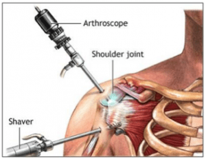
A brief history on the evolution of shoulder arthroscopy (Dessai, 2020):
The invention of the incandescent light bulb by Edison in 1879, led to the introduction of the laparo-thoracoscope in 1910. Attempts were made to use this device in the knee joint as well. Development of the arthroscope really took off after the introduction of “cold-light” and rod lens optical system by Hopkins in 1960. Kenji Takagi and later Masaki Watanabe get the credit for developing the modern form of arthroscopy.
The spillover of knee arthroscopy into the shoulder was inevitable and began in the 1980s. Shoulder arthroscopy started with instability repair, followed by subacromial decompression (relieving the pressure between the bone on top of the shoulder joint and the top part of the upper arm bone). Through the 1980s and 1990s, with developments in biotechnology, more sophisticated tools and anchors became available, leading to refinement of instability repair procedures.
Remember, arthroscopic surgery is not a panacea and certainly no substitute for a good clinical evaluation.
Total Shoulder Replacement surgery:
The first Total Shoulder Replacement surgery was performed in 1893 by the French surgeon Jules Emil Pean, as treatment for a 37-year-old baker dying of a tuberculosis infection in his (R) shoulder. Pean used a platinum and rubber implant in the patient that lasted 2 years and allowed the young man to return to his work.
The next generation of prostheses came in the 1950s, with the advent of the plastic and acrylic humeral head (the ball part of the joint). The implants were typically fixed with plates and screws farther down the humerus (upper arm bone) and failed due to the screws being pulled out of the bone. Several iterations of implant designs and materials appeared through the early 1980s, again, with limited success. By the 1990s, new designs resulted in marked functional improvements and consistently more positive outcomes. Currently, up to 90% of shoulder replacements last for 10 years or more.
As with the knee, what I frequently hear from patients is; ‘My X-ray is bone on bone,’ or, ‘I was told that ‘this is the worst I’ve ever seen,’ or ‘according to your MRI, surgery is the only way you will have the use of your arm.’
Again, the image should not make the diagnosis. It is just one piece of a multi-factorial presentation, including, but not limited to current function and mobility, the patient’s lifestyle and needs, risk/benefit analysis, recovery time, patient age and health status, each to be carefully considered. The surgery-decision-making criteria remain the same for the shoulder as in the knee: you are no longer able to perform your daily activities and/or your shoulder is so painful that you are unable to function.
Understanding Frozen Shoulder:
Any discussion of shoulder dysfunction and its treatment must include frozen shoulder, a musculoskeletal condition unique to the shoulder. According to Griggs et al (2000), the largest single group of patients have no detectable underlying cause for their symptoms (primary idiopathic frozen shoulder). A substantial group of patients with diabetes mellitus (Type II Diabetes) are considered as a separate group, Diabetic Frozen Shoulder, since the course is usually more severe (with regard to pain and limitation and tend to be more protracted (lasting much longer), presumably due to compromised circulation common in diabetes.
Patients with frozen shoulder will report that the symptoms come on spontaneously, without any apparent cause. Radiologic findings are frequently negative and the symptoms are rarely associated with pathologic findings. The diagnosis is made with the history and clinical presentation:
The three phases of frozen shoulder:
- Initial phase: characterized by significant should pain with movement and/or at rest
- Freezing phase: characterized by a decrease in pain with progressive limitation in shoulder elevation and rotational motions
- Thawing phase: characterized by progressive increase in range of motion and function.
Surgery is rarely an option with frozen shoulder. Occasionally, manipulation under anesthesia is performed, where the patient is put to sleep and the shoulder is forcibly moved. This procedure typically results in a worsening of the symptoms and a more protracted course. Physical therapy is typically the treatment of choice. With no intervention, the typical course is 18 months to resolution. With appropriate physical therapy, the length of the clinical course can be significantly shortened (6-9 months).
|