|
Background
Ethylene glycol is a toxic alcohol characterized by a low freezing point, which underlies its widespread use across both household and industrial applications. It is a common constituent of automotive antifreeze, radiator coolant, and de-icing solutions, and is also utilized as a solvent in paints, inks, and resins; it may additionally be found in products such as snow globes. “Pet-safe” antifreeze formulations often substitute propylene glycol for ethylene glycol.
Ethylene glycol is a colorless, odorless liquid with an inherently sweet taste, a property that contributes to its risk for unintentional ingestion. According to 2024 data from U.S. poison centers, there were 631 reported single-substance ethylene glycol exposures and 7,079 additional cases in which ethylene glycol was involved as part of a multi-substance exposure (Beuhler et al., 2025). Toxicity most commonly occurs via oral ingestion.
Pathophysiology
Ethylene glycol toxicity is mediated by both its direct central nervous system (CNS) depressant properties and the generation of toxic metabolites following hepatic biotransformation. Clinical effects are classically described across three domains: CNS depression, cardiopulmonary compromise, and renal injury. Post ingestion, ethylene glycol is rapidly absorbed from the gastrointestinal tract, with peak serum concentrations occurring within a short interval.
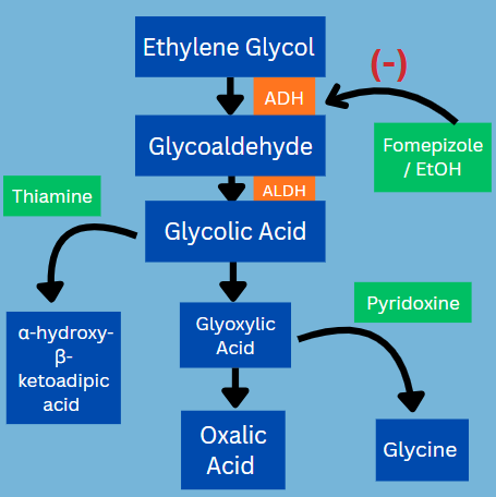
Hepatic metabolism occurs via alcohol dehydrogenase to glycolaldehyde, which is subsequently oxidized to glycolic acid. Glycolic acid is the primary driver of high-anion-gap metabolic acidosis (AGMA) and is associated with elevated measured lactate. Further metabolism yields oxalic acid, which chelates calcium to form insoluble calcium oxalate crystals, resulting in tissue deposition and acute kidney injury. In the absence of prompt intervention, accumulation of these metabolites may persist for several days, thereby prolonging toxicity and increasing the risk of end-organ damage (Scalley et al. 2002).
Clinical toxicity and evaluation
The clinical course of ethylene glycol poisoning characteristically evolves through temporally defined phases. During the initial 12 hours following ingestion, patients commonly demonstrate central nervous system depression with inebriation-like features, including euphoria. If the patient has not coingested ethanol, they may start with an AGMA early. Between 12 and 24 hours, the progressive accumulation of acidic metabolites leads to AGMA, which is frequently accompanied by compensatory hyperventilation. By 48 to 72 hours post-ingestion, renal manifestations predominate, including oliguria and flank pain, reflecting acute kidney injury secondary to calcium oxalate crystal deposition. Hypocalcemia, although rare, may result from calcium chelation, and intrarenal crystal deposition can contribute to obstructive phenomena such as hydronephrosis (Hodgman et al. 2017). Additional clinical findings may include tachycardia, cardiac dysrhythmias, electrolyte abnormalities, and gastrointestinal symptoms, including nausea and vomiting (Head et al. 2012).
The diagnostic gold standard for ethylene glycol poisoning is direct measurement of the serum or plasma ethylene glycol concentration. However, this test is not always rapidly available and does not reliably correlate with clinical severity. In Ohio, several laboratories can perform ethylene glycol testing, and clinicians should promptly contact their regional poison control center for assistance to locate them. Every effort should be made to obtain a level as quickly as possible.
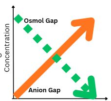
Adjunctive laboratory evaluation and calculating the osmolar gap and anion gap can provide useful diagnostic clues, but both are prone to error and must be interpreted cautiously (Ralph et al. 2025). Early in toxicity, the osmolar gap is often elevated due to unmetabolized ethylene glycol; as metabolism progresses and toxic organic acids accumulate, the osmolar gap may normalize. In contrast, the anion gap typically rises later, reflecting the development of a high AGMA.
Although serum ethylene glycol concentration can be estimated from the osmolar gap using a conversion factor of approximately 6.2, this approach has limitations, particularly in the presence of co-ingested ethanol, which confounds interpretation. Clinically, the combination of an elevated osmolar gap and a high AGMA should raise a strong suspicion for toxic alcohol exposure.
Treatment
Fomepizole is the preferred antidote for ethylene glycol poisoning due to its potent and selective inhibition of alcohol dehydrogenase, thereby preventing the formation of toxic metabolites. In settings where fomepizole is unavailable, ethanol may be employed as an alternative competitive substrate for alcohol dehydrogenase; however, its use necessitates intensive monitoring given its central nervous system depressant effects and narrow therapeutic index. Antidotal therapy is generally indicated in patients with elevated serum ethylene glycol concentrations (commonly >20–25 mg/dL) or in the presence of clinical or laboratory evidence of metabolic acidosis and/or renal dysfunction.
Hemodialysis should be considered in patients with severe metabolic acidosis, electrolyte abnormalities, renal failure, or markedly elevated serum ethylene glycol concentrations, as it effectively removes both the parent compound and its toxic metabolites while correcting acid–base derangements. Adjunctive therapy includes thiamine (vitamin B1) and pyridoxine (vitamin B6), which facilitate alternative metabolic pathways promoting the conversion of glyoxylic acid to non-toxic metabolites, including glycine and α-hydroxy-β-ketoadipate, respectively. Supportive care should be individualized according to the severity of toxicity and overall clinical trajectory (Scalley et al. 2002).
|